Copyright
©The Author(s) 2017.
World J Cardiol. Jul 26, 2017; 9(7): 629-633
Published online Jul 26, 2017. doi: 10.4330/wjc.v9.i7.629
Published online Jul 26, 2017. doi: 10.4330/wjc.v9.i7.629
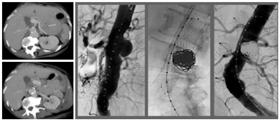
Figure 1 AngioCT scan and digital substraction angiography.
Mycotic aneurysm and the relation with the visceral branches, stent assisted coil embolization of aneurysms with a Sinus XL stent and complete embolization of the mycotic aneurysms without evidence of flow inside the sac.
Figure 2 Digital substraction angiography (A-C).
Flow in the mycotic aneurysms an increased de diameter of the sac. Coils and n-butyl 2-cyanoacrylate embolization performed with the balloon-assisted technique and final angiographic control shows absence of flow in the interior of the aneurysms.
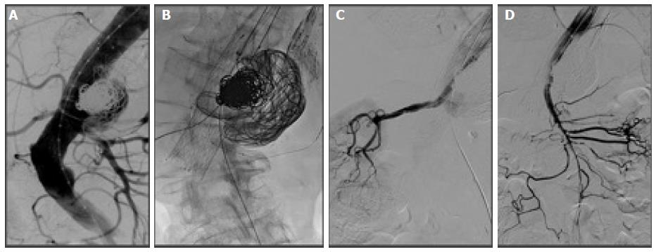
Figure 3 Digital substraction angiography.
Rechanneling of the pseudoaneurysms. Renals and superior mesenteric arteries catheterization (the left renal artery with a coronary balloon angioplasty) and the presence of n- butyl 2-cyanoacrylate out of the pseudoaneurysms. Chimney in the right renal artery and superior mesenteric artery.
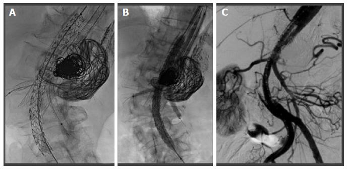
Figure 4 Single Nellix endovascular sealing and the chimneys (A-C).
Fluoroscopy of the balloons inflated during the filling of the bag of the Nellix EVAS with the polymer. Permeability of the endograft, both chimneys and the celiac trunk without evidence of endoleaks.
- Citation: Rabellino M, Moltini PN, Di Caro VG, Chas JG, Marenchino R, Garcia-Monaco RD. Endovascular treatment of paravisceral mycotic aneurysm: Chimmeny endovascular sealing the end of de road. World J Cardiol 2017; 9(7): 629-633
- URL: https://www.wjgnet.com/1949-8462/full/v9/i7/629.htm
- DOI: https://dx.doi.org/10.4330/wjc.v9.i7.629


