Copyright
©The Author(s) 2017.
World J Cardiol. Jun 26, 2017; 9(6): 558-561
Published online Jun 26, 2017. doi: 10.4330/wjc.v9.i6.558
Published online Jun 26, 2017. doi: 10.4330/wjc.v9.i6.558
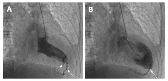
Figure 1 Inadvertent phlebography of the posterior interventricular vein.
Right anterior oblique image taken at time of left ventriculography. Arrows show the posterior interventricular vein in systole (A) and diastole (B). Arrowhead shows a minor subendocardial staining (A).
- Citation: Aznaouridis K, Masoura C, Kastellanos S, Alahmar A. Inadvertent cardiac phlebography. World J Cardiol 2017; 9(6): 558-561
- URL: https://www.wjgnet.com/1949-8462/full/v9/i6/558.htm
- DOI: https://dx.doi.org/10.4330/wjc.v9.i6.558


