Copyright
©The Author(s) 2025.
World J Cardiol. Jun 26, 2025; 17(6): 106445
Published online Jun 26, 2025. doi: 10.4330/wjc.v17.i6.106445
Published online Jun 26, 2025. doi: 10.4330/wjc.v17.i6.106445
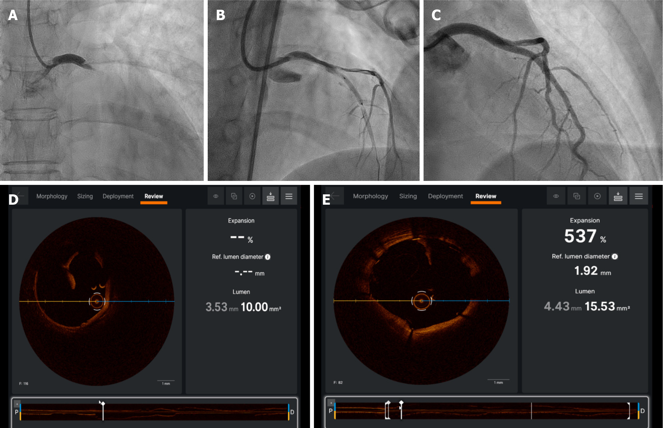
Figure 1 A 25-year-old postpartum female who presented with extensive anterior ST-elevation myocardial infarction and found to have spontaneous coronary artery dissection involving left main, left anterior descending artery and left circumflex artery.
A: It shows distal left main (LM) dissection and left anterior descending artery (LAD)/left circumflex artery (LCX) total occlusion; B: It shows wiring of LAD/LCX; C: It shows post percutaneous coronary intervention to LM/LAD- LCX; D and E: They show optical coherence tomography imaging showing LM/LAD dissection.
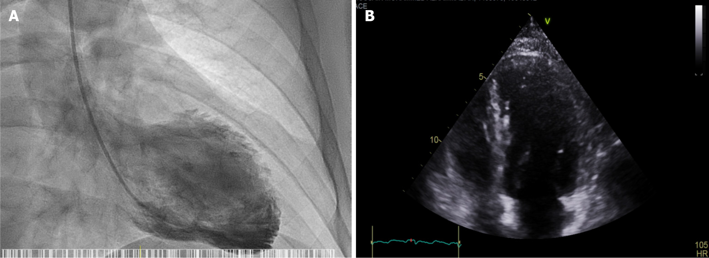
Figure 2 Left Ventricle Angiogram.
A: It shows Left Ventricle Angiogram showing apical ballooning; B: Echocardiogram shows Apical ballooning.
- Citation: Hegazi Abdelsamie A, Abdelhadi HO, Abdelwahed AT. Acute myocardial infarction in the young: A 3-year retrospective study. World J Cardiol 2025; 17(6): 106445
- URL: https://www.wjgnet.com/1949-8462/full/v17/i6/106445.htm
- DOI: https://dx.doi.org/10.4330/wjc.v17.i6.106445


