Copyright
©The Author(s) 2022.
World J Cardiol. Sep 26, 2022; 14(9): 496-507
Published online Sep 26, 2022. doi: 10.4330/wjc.v14.i9.496
Published online Sep 26, 2022. doi: 10.4330/wjc.v14.i9.496
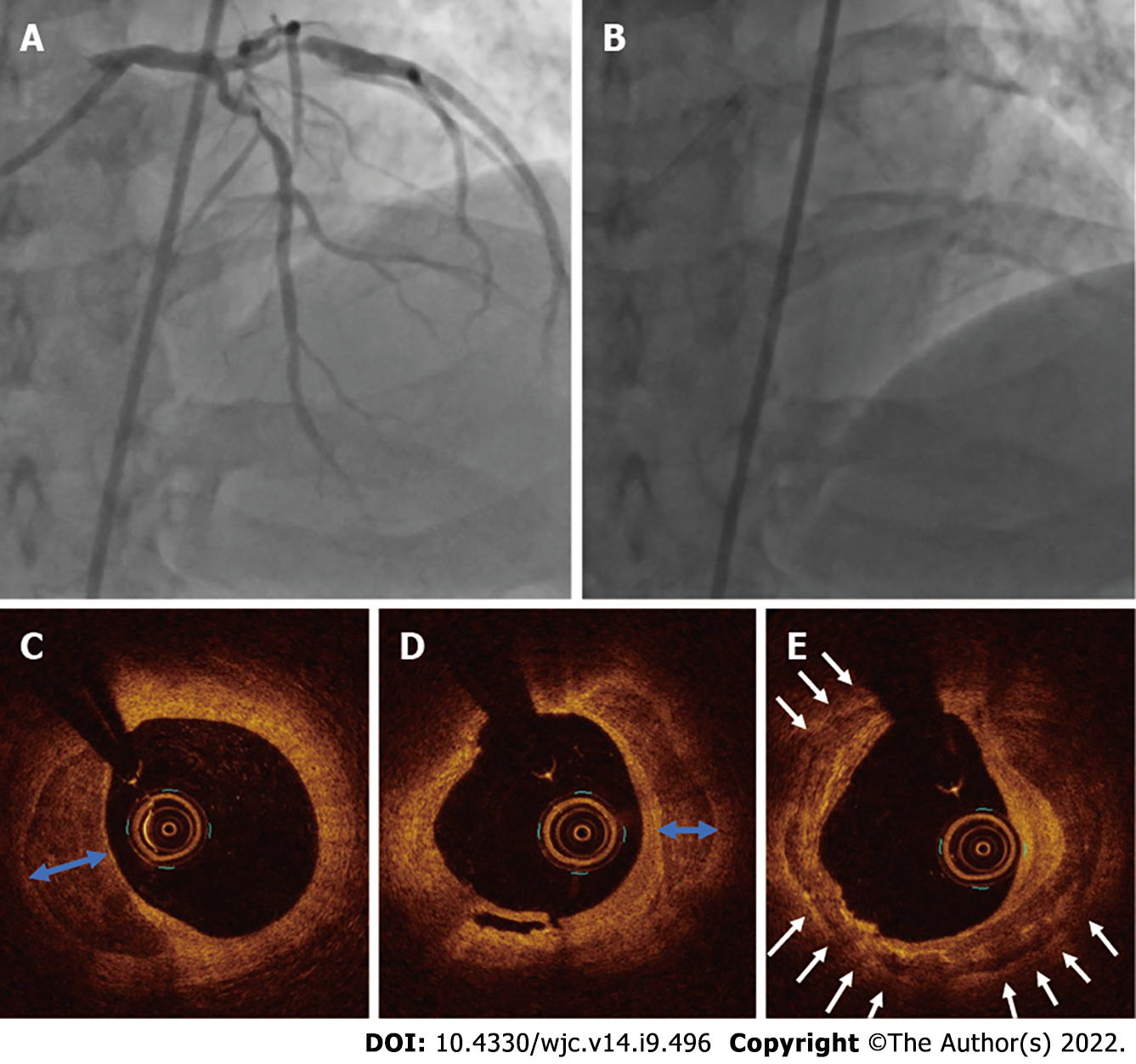
Figure 1 Coronary angiogram of case 1.
A and B: A severe calcific lesion in the left anterior descending coronary artery and proximal major obtuse marginal artery; C-E: An optical coherence tomography showed circumferential (white arrows) and deep calcium arc (blue arrow) prior to percutaneous coronary intervention.
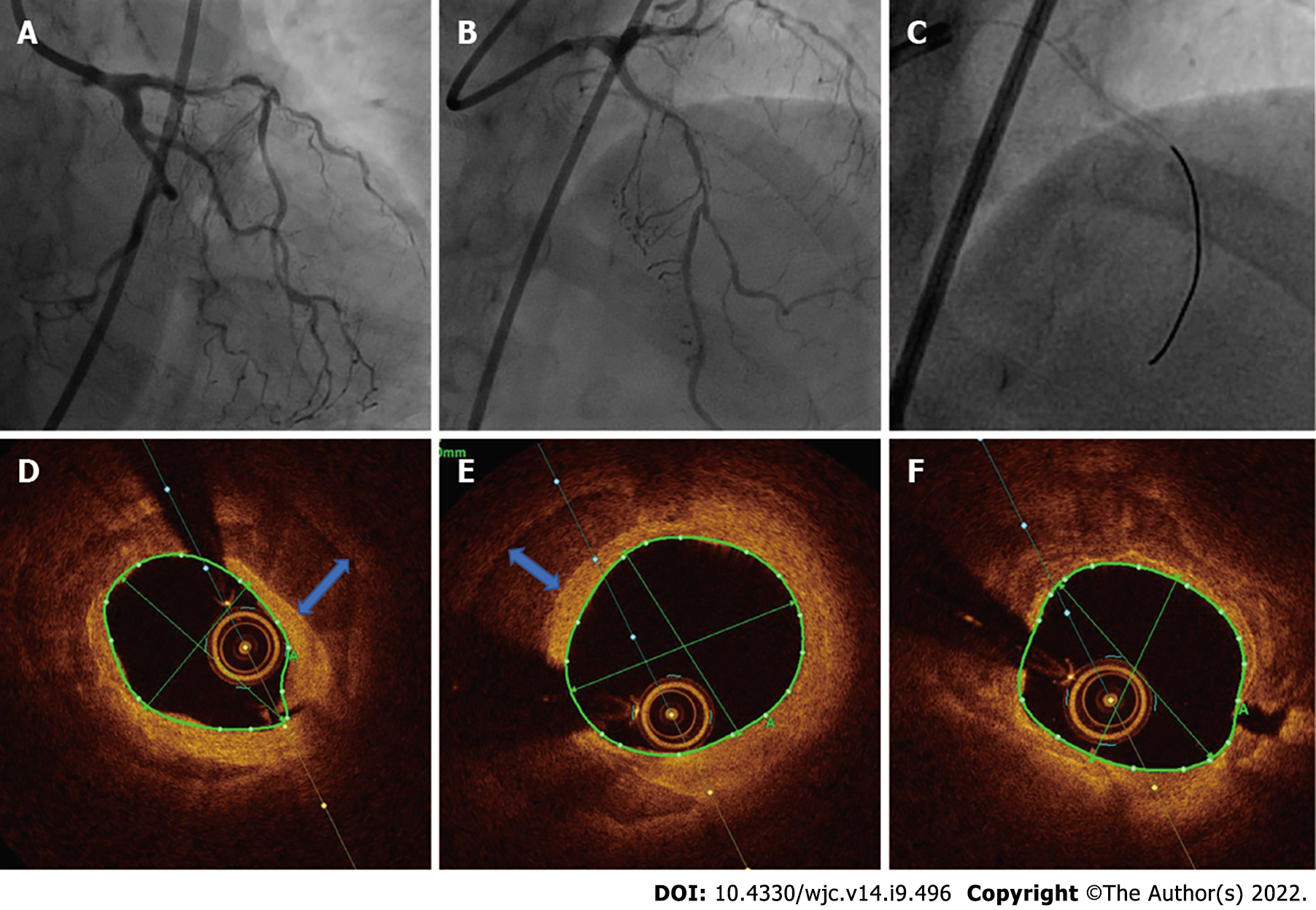
Figure 2 Coronary angiogram of case 2.
A-C: A severe calcific lesion in the left anterior descending coronary artery; D-F: Optical coherence tomography showed circumferential calcium and deep calcium (blue arrow) prior to percutaneous coronary intervention.
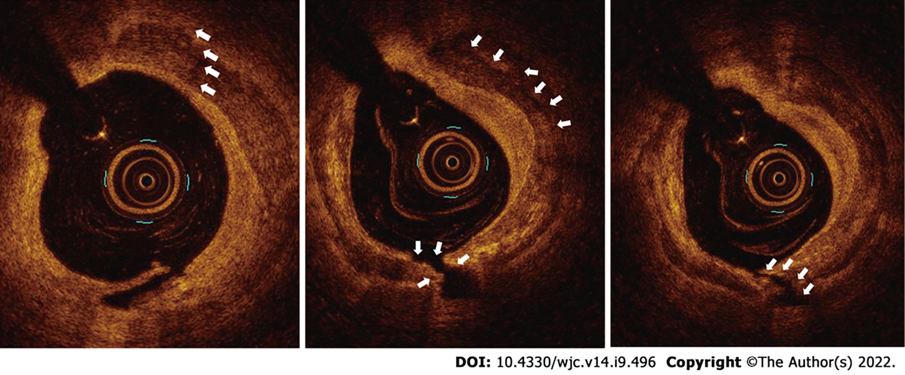
Figure 3 Post intravascular lithotripsy optical coherence tomography images of left anterior descending coronary artery of case 1 depicting calcium fracture (white arrow).
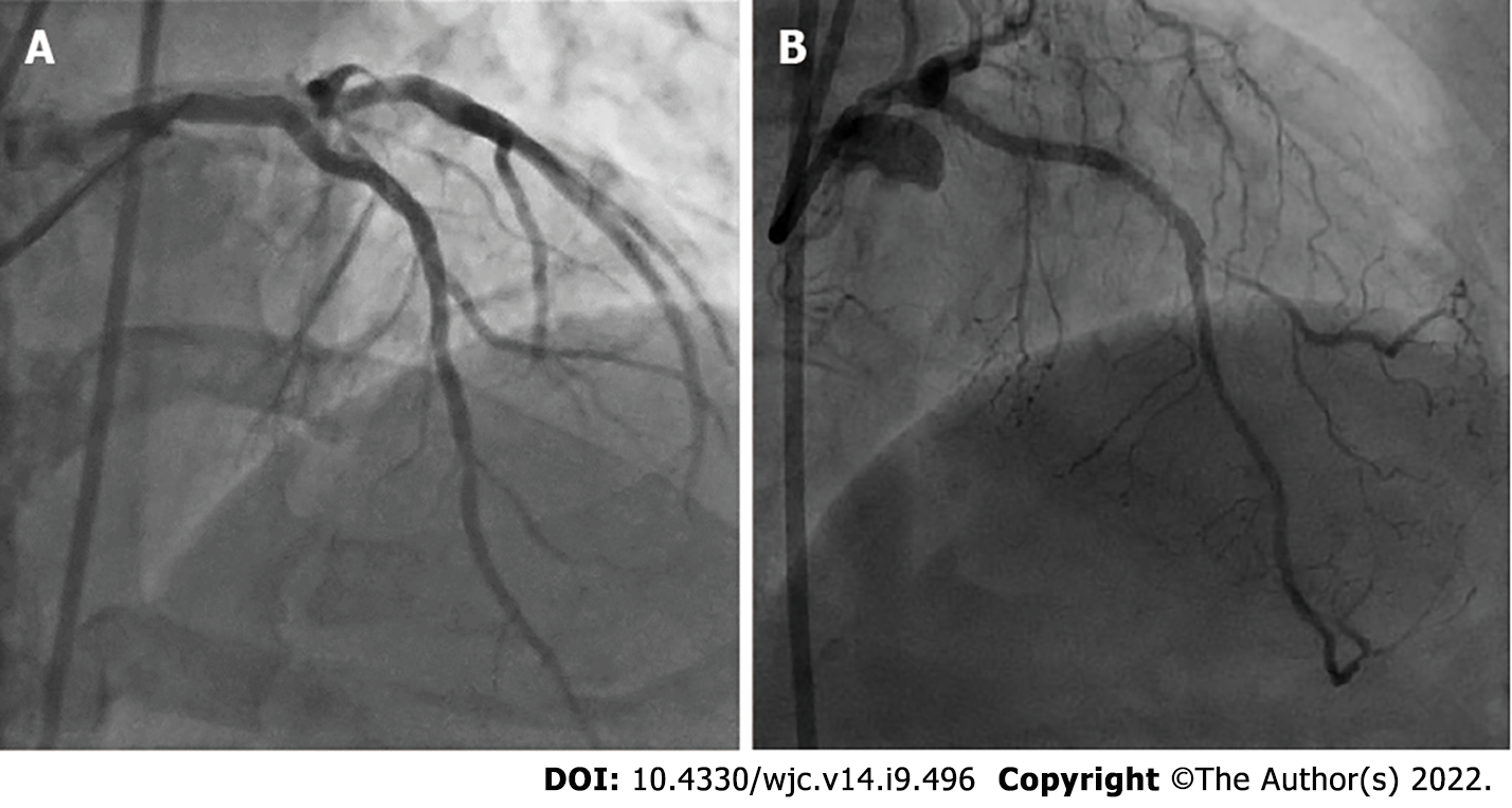
Figure 4 Post percutaneous coronary intervention coronary angiogram.
A: Post percutaneous coronary intervention coronary angiogram showed a well expanded left anterior descending coronary artery in case 1; B: Post percutaneous coronary intervention coronary angiogram showed fully expanded stent and thrombolysis in myocardial infarction 3 flow in case 2.
- Citation: Pradhan A, Vishwakarma P, Bhandari M, Sethi R. Intravascular lithotripsy for coronary calcium: A case report and review of the literature. World J Cardiol 2022; 14(9): 496-507
- URL: https://www.wjgnet.com/1949-8462/full/v14/i9/496.htm
- DOI: https://dx.doi.org/10.4330/wjc.v14.i9.496


