Copyright
©The Author(s) 2020.
World J Biol Chem. Sep 27, 2020; 11(2): 30-51
Published online Sep 27, 2020. doi: 10.4331/wjbc.v11.i2.30
Published online Sep 27, 2020. doi: 10.4331/wjbc.v11.i2.30
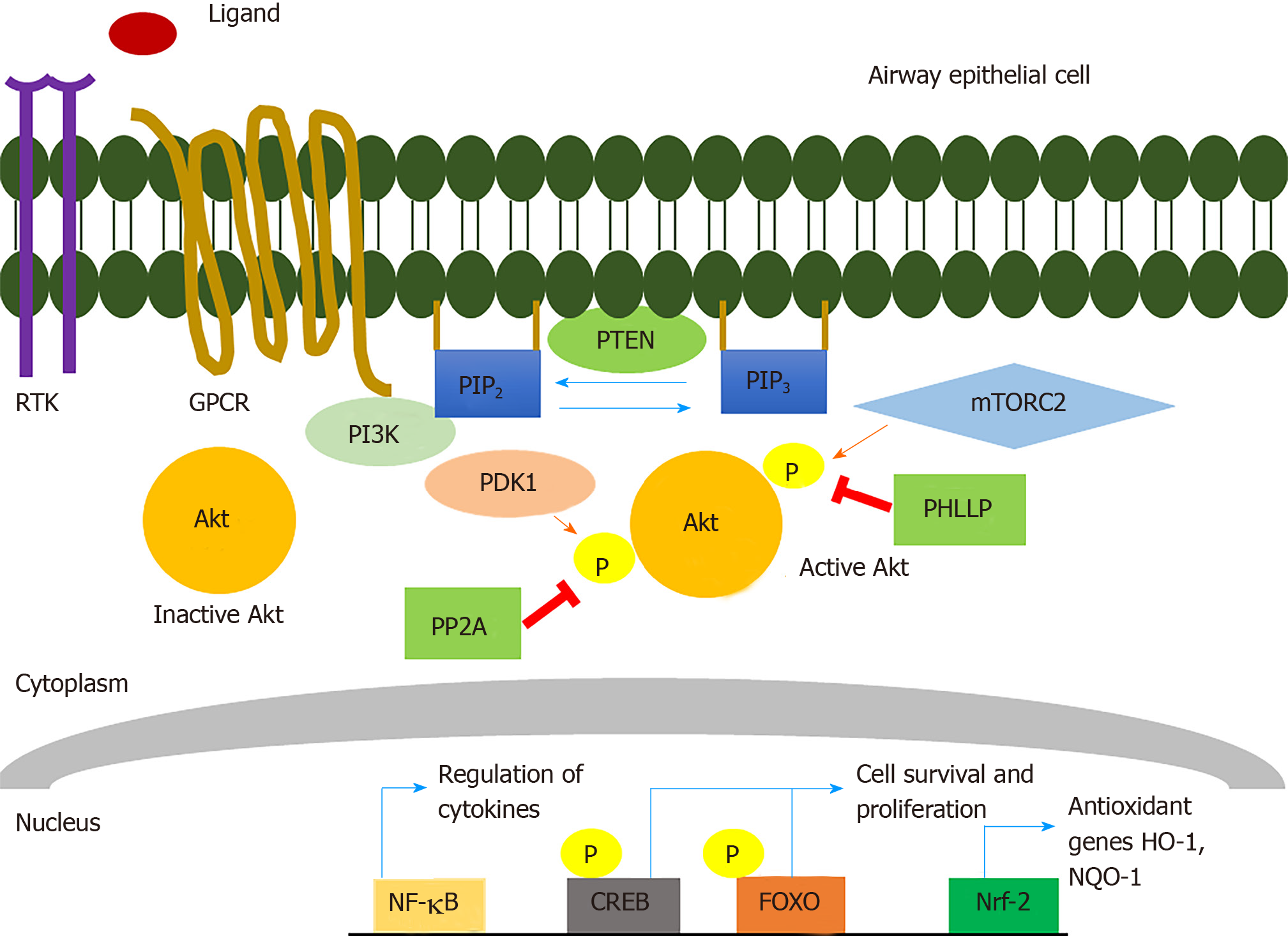
Figure 1 Protein kinase B signaling pathway.
Stimulation of receptor tyrosine kinases or G-protein-coupled-receptors activates phosphatidylinositol (3,4,5)-trisphosphate (PIP3) which phosphorylates phosphatidylinositol 4,5-bisphosphate (PIP2) at the plasma membrane to generate PIP3. Inactive Akt in the cytosol gets recruited to the plasma membrane where it gets phosphorylated at T308 in the kinase domain by phosphoinositide dependent kinase 1 and at S473 in the regulatory domain by mTORC2 resulting in full activation; Signal termination is achieved by PTEN where it dephosphorylates PIP3 to PIP2; Additionally, PP2A and PHLPP have shown to regulate Akt kinase activity by direct dephosphorylation; Activation of Akt is known to regulate crucial transcription factors such as nuclear factor-kB (NF- kB), CREB, FOXO, and Nrf-2, each of which regulates a variety of target genes that regulate cell survival, proliferation, differentiation, migration, and metabolism; Akt is known to phosphorylate IkB kinase which phosphorylates IkB-a releasing NF-kB to translocate into the nucleus and transcribe genes; Activation of Akt might increase or suppress NF-kB regulated genes (IL-8, IL-18) depending on the stimulus. Both FOXO and CREB are known to regulate apoptosis and phosphorylation of these transcription factors by Akt, has shown to control cell survival. Nrf-2 activation by Akt increases the production of antioxidant genes such as HO-1 and NQO-1 that counteracts oxidative stress and inflammation. RTK: Receptor tyrosine kinase; GPCR: G-protein-coupled receptor; PI3K: Phosphoinositide-3-kinase; PIP2: Phosphatidylinositol 4,5-bisphosphate; Akt: Protein kinase B; PDK1: Phosphoinositide dependent kinase 1; mTORC2: Mammalian target of rapamycin; PTEN: Phosphatase and tensin homolog; PHLPP: PH domain and leucine rich repeat protein phosphatases; NF-kB: Nuclear factor kappa-light-chain-enhancer of activated B cells; CREB: cAMP response element binding protein; FOXO: Forkhead family of transcription factors; Nrf-2: Nuclear factor erythroid 2 related factor-2; PP2A: Protein phosphatase 2; HO-1: Heme oxygenase; NQO-1: NADPH quinone dehydrogenase 1.
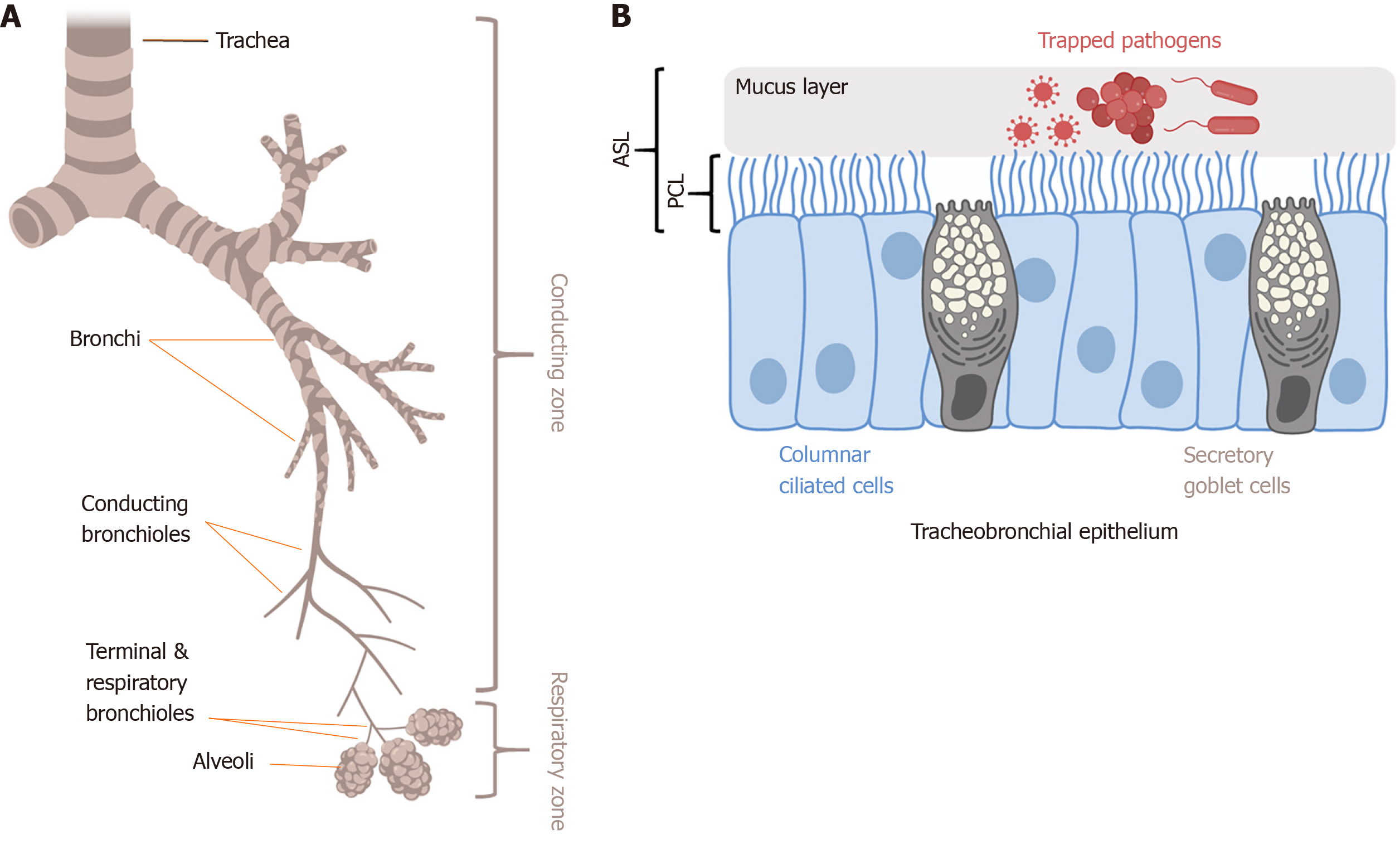
Figure 2 Mucociliary clearance and innate immunity in the lung.
A: Trachea, bronchi, and conducting bronchioles comprise the conducting zone of the airways; B: The conducting airway epithelium is lined with columnar motile ciliated cells and secretory goblet cells. Goblet cells secrete mucins like MUC5AC that polymerize to form mucus, which traps inhaled pathogens and debris; The mucus layer rides on top of a less viscous PCL composed of salt, water, and antimicrobials; Together, the mucus and PCL comprise the airway surface liquid; Coordinated metachronal beating of the motile cilia within the PCL layer pushes the sticky mucus layer up to respiratory tree to the oropharynx, where it is expectorated or swallowed; This process is termed MCC, and is the physical defense of the airway against infection; Epithelial cells also secrete antimicrobial peptide and radicals (NO, H2O2) to directly kill pathogens and produce cytokines and chemokines to activate inflammation; Shown is a representative diagram of tracheal or bronchial epithelium; In lower conducting airways (non-cartilaginous bronchioles < approximately 1 mm in diameter), secretory club cells (also known as bronchiolar exocrine cells) are found instead of goblet cells; As described in the text, there are several potential mechanisms by which protein kinase B may regulate MCC and other innate immune pathways. Figure made using Biorender.com. PCL: Periciliary layer; MCC: Mucociliary clearance; MUC5AC: Mucin 5AC.
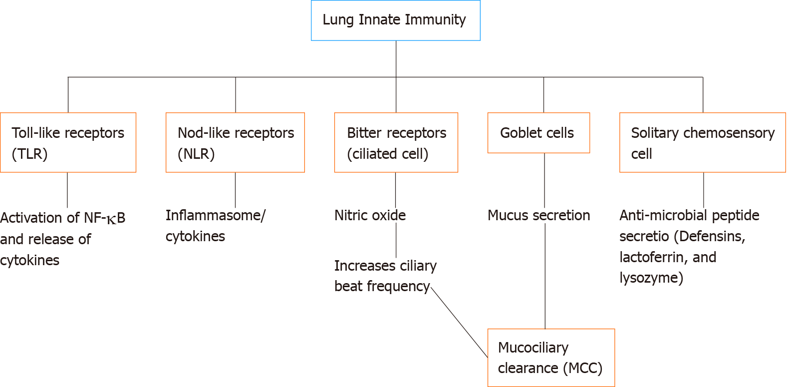
Figure 3 Summarization of non-specific immune strategies in lung innate immunity.
Toll-like receptors are activated to recognize pathogen-associated microbial patterns/damage-associated molecular pattern (PAMPs/DAMPs) and activate nuclear factor-kB to produce cytokines; nod-like receptors are cytosolic receptors that can recognize intracellular PAMPs/DAMPs and can form inflammasome to either produce cytokines such as interferons or induce pyroptosis (inflammation associated apoptosis); Stimulation of bitter receptors, expressed in ciliated cells in the airways, elevates Ca2+ that produces NO which activates protein kinase G and increase ciliary beat frequency; NO can directly kill bacteria; Goblet cells produce mucus that traps pathogens and bacteria which are then eliminated out of the upper airway system through MCC; Activation of bitter receptors in solitary chemosensory cells can increase intracellular Ca2+ that can produce antimicrobial peptides.
- Citation: Gopallawa I, Lee RJ. Targeting the phosphoinositide-3-kinase/protein kinase B pathway in airway innate immunity. World J Biol Chem 2020; 11(2): 30-51
- URL: https://www.wjgnet.com/1949-8454/full/v11/i2/30.htm
- DOI: https://dx.doi.org/10.4331/wjbc.v11.i2.30


