Published online Oct 27, 2013. doi: 10.4240/wjgs.v5.i10.264
Revised: September 11, 2013
Accepted: October 16, 2013
Published online: October 27, 2013
Processing time: 89 Days and 17 Hours
Cystic lymphangiomas are rare benign tumors. Most frequently occurring in children and involving the neck or axilla, these tumors are much less common in adults and very rarely involve the abdomen. The known congenital and acquired (traumatic) etiologies result in failure of the lymphatic channels and consequent proliferation of lymphatic spaces. This case report describes a very rare case of a giant mesenteric cystic lymphangioma in an adult male with no clear etiology and successful resolution by standard radical resection. A previously healthy 44-year-old male presented with a 6-wk history of progressive upper abdominal pain, vomiting, anorexia and unintentional weight loss accompanied by rapid abdominal distension. A palpable mass was detected upon physical examination of the distended abdomen and abdominal computed tomography scan showed a giant multilobulated cystic process, measuring 40 cm in diameter. Exploratory laparotomy revealed an enormous cystic mass containing 6 L of serous fluid. The process appeared to originate from the lesser omentum and the lesser curvature of the stomach. Radical resection of the tumor was performed along with a partial gastrectomy to address potential invasion into the adjacent tissues. Histological analysis confirmed the diagnosis of a multicystic lymphangioma. The postoperative recovery was uneventful and the patient was discharged after 6 d. At 3-mo follow-up, the patient was in good health with no signs of recurrence.
Core tip: A 44-year-old man presented with abdominal distension and unexplained weight loss. Computed tomography scan of the abdomen showed an enormous multilobulated cystic process. Subsequent exploratory laparotomy revealed a cystic mass containing 6 L serous fluid, originating from the lesser curvature of the stomach. Pathological examination of the resected specimen obtained by radical partial gastrectomy indicated multicystic lymphangioma, a rare benign tumor caused by congenital or traumatic defects of lymphatic channels. Although these large tumors can cause mass effect symptoms and result in serious complications, this case had an uneventful recovery and no complaints at 3-mo follow-up.
- Citation: van Oudheusden TR, Nienhuijs SW, Demeyere TBJ, Luyer MDP, de Hingh IHJT. Giant cystic lymphangioma originating from the lesser curvature of the stomach. World J Gastrointest Surg 2013; 5(10): 264-267
- URL: https://www.wjgnet.com/1948-9366/full/v5/i10/264.htm
- DOI: https://dx.doi.org/10.4240/wjgs.v5.i10.264
Cystic lymphangiomas are rare, slow-growing, benign tumors, originating from lymphatic channels[1]. Most frequently, these tumors are diagnosed in children (up to 90% manifest before the age of three[2-4]) and are generally localized to the neck or axilla (95% of cases); localization to the abdominal cavity is more common among the rare adult cases[5,6]. Regardless of age, clinical presentation is diverse, varying from complaints of mild nausea to acute abdominal pain, with mass effect accounting for the transition to a symptomatic state[7]. Diagnosis in asymptomatic cases has been made as a coincident finding during laparotomies to address other issues[8].
Here, we describe the case of a giant mesenteric cystic lymphangioma (GMCL) occurring in a middle-aged adult male, which was successfully treated with partial gastrectomy. At > 44 cm, this GMCL is the largest reported to date in the publicly available literature.
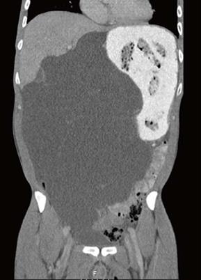
A 44-year-old man presented to a local clinic with a roughly 6-wk history of progressive upper abdominal pain, vomiting, and anorexia. The patient also reported unintentional weight loss of at least 4 kg that was accompanied by a progressive distension of the abdomen. Upon referral to our center for further analysis, computed tomography (CT) was performed to evaluate the palpable abdominal mass detected by routine physical examination. The CT-scan showed a giant multilobulated cystic process, measuring at least 40 cm in diameter (Figure 1), with unclear origin. The differential diagnosis at that time contained cystic lymphangioma, duplication cyst, pancreatic cyst, gastro-intestinal stromal tumor and multicystic mesothelioma.
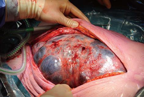
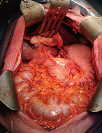
During this initial examination period, the patient could not tolerate any food and required continuous analgesic drug treatment to control severe abdominal pain. An exploratory laparotomy was performed and revealed an enormous lobulated mass at the midline incision (Figure 2). Near complete drainage of the mass (total of 6 L serous fluid) was required to gain access to the abdominal cavity. The tumor was then observed to originate from the lesser omentum and lesser curvature of the stomach. The left gastric artery and vein were found to be completely enveloped by the tumor, and were therefore dissected. Radical resection of the tumor along the minor curvature of the gastric antrum was carried out with a tri-stapler (Figure 3).
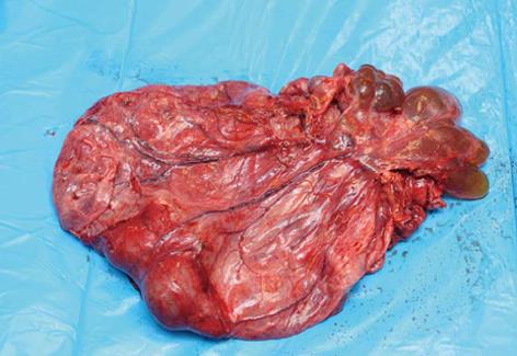
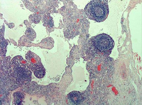
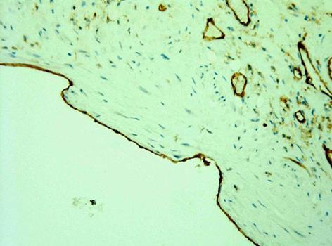
The resected mass process, with some remaining fluid, measured 19 cm × 17 cm × 4 cm (Figure 4). Pathological analysis confirmed the diagnosis of multicystic lymphangioma attached to the serosal side of the stomach, and indicated no signs of malignancy. Diagnosis was based on dilated cystic structures (Figure 5), delineated with endothelial lining as proven by CD31-staining (Figure 6). Absence of red blood cells and presence of lymphoid tissue confirmed the diagnosis of lymphangioma. Postoperative recovery was uneventful and the patient was discharged 6 d later with complete relief of all prior symptoms. At 3-mo follow-up, the patient appeared in good health and had no complaints.
The precise etiology of cystic lymphangiomas remains to be fully elucidated, but involves acquired or congenital failure of lymphatic channels. While the latter may zmore childhood manifestations, the former is more likely associated with the adult manifestations, possibly related to inflammatory conditions or physical trauma such as created by surgical or radiation therapies[3,9]. These two etiologies are not mutually exclusive, and it is possible that a congenital impairment in communication between mesenchymal slits and the venous system may put a patient at greater risk of drainage blocking in response to trauma[10]. However, in the current adult case described herein, no precipitating cause could be identified.
Mesenteric lymphangiomas are histologically distinct from mesenteric cysts. While mesenteric cysts originate from mesothelial tissue[11], lymphangiomas are composed of alternating lymphoid tissue, lymphatic space, and foam cells. Thus, the histological analysis of the resected mass from our patient provided a clear result for this differential diagnosis.
The presenting symptoms of the current case were in line with the characteristic mass effect symptomology of cystic lymphangiomas, including nausea, weight loss and abdominal distension[12]. However, as the mass size reaches a critical threshold, and depending on its spatial orientation, more severe and life-threatening complications may occur, such as partial bowel obstruction, torsion, volvulus, perforation or rupture, and hemoperitoneum. In some cases, nerves are entrapped in the large mass, resulting in additional pain[13-15]. Therefore, immediate treatment is recommended upon diagnosis.
The treatment of choice is surgical resection, usually carried out by laparoscopy which has good safety and efficacy profiles[1,4,15]. The particularly enormous size of the cystic lymphangioma in our case further justified use of the laparotomy approach, since no free space was present in the abdomen. In addition, a radical resection (involving tissues of the related organ of origin) was performed to help reduce the risk of recurrence since these tumors, although benign, tend to invade adjacent structures[11].
In conclusion, this case report describes a rare adult case of cystic mesenteric lymphangioma. Despite being the largest tumor of its type reported to date, the standard surgical treatment of radical resection including a partial gastrectomy was able to rapidly and successfully resolve the condition. Cystic mesenteric lymphangioma should be considered as differential diagnosis when patients present with a multilobulated cystic tumor in the abdomen. Radical resection remains a sufficiently safe and effective treatment option for this type of benign tumor.
P- Reviewers Kim WH, Pavlidis TE S- Editor Zhai HH L- Editor A E- Editor Lu YJ
| 1. | Losanoff JE, Richman BW, El-Sherif A, Rider KD, Jones JW. Mesenteric cystic lymphangioma. J Am Coll Surg. 2003;196:598-603. [PubMed] |
| 2. | Alqahtani A, Nguyen LT, Flageole H, Shaw K, Laberge JM. 25 years’ experience with lymphangiomas in children. J Pediatr Surg. 1999;34:1164-1168. [RCA] [PubMed] [DOI] [Full Text] [Cited by in RCA: 1] [Reference Citation Analysis (0)] |
| 3. | Rieker RJ, Quentmeier A, Weiss C, Kretzschmar U, Amann K, Mechtersheimer G, Bläker H, Herwart OF. Cystic lymphangioma of the small-bowel mesentery: case report and a review of the literature. Pathol Oncol Res. 2000;6:146-148. [RCA] [PubMed] [DOI] [Full Text] [Cited by in Crossref: 41] [Cited by in RCA: 51] [Article Influence: 2.0] [Reference Citation Analysis (0)] |
| 4. | Fernández Hurtado I, Bregante J, Mulet Ferragut JF, Morón Canis JM. [Abdominal cystic lymphangioma]. Cir Pediatr. 1998;11:171-173. [PubMed] |
| 5. | Hebra A, Brown MF, McGeehin KM, Ross AJ. Mesenteric, omental, and retroperitoneal cysts in children: a clinical study of 22 cases. South Med J. 1993;86:173-176. [RCA] [PubMed] [DOI] [Full Text] [Cited by in Crossref: 73] [Cited by in RCA: 69] [Article Influence: 2.2] [Reference Citation Analysis (0)] |
| 6. | Roisman I, Manny J, Fields S, Shiloni E. Intra-abdominal lymphangioma. Br J Surg. 1989;76:485-489. [RCA] [PubMed] [DOI] [Full Text] [Cited by in Crossref: 118] [Cited by in RCA: 121] [Article Influence: 3.4] [Reference Citation Analysis (0)] |
| 7. | Rami M, Mahmoudi A, El Madi A, Khalid MA, Bouabdallah Y. Giant cystic lymphangioma of the mesentery: varied clinical presentation of 3 cases. Pan Afr Med J. 2012;12:7. [PubMed] |
| 8. | de Vries JJ, Vogten JM, de Bruin PC, Boerma D, van de Pavoordt HD, Hagendoorn J. Mesenterical lymphangiomatosis causing volvulus and intestinal obstruction. Lymphat Res Biol. 2007;5:269-273. [RCA] [PubMed] [DOI] [Full Text] [Cited by in Crossref: 7] [Cited by in RCA: 11] [Article Influence: 0.6] [Reference Citation Analysis (0)] |
| 9. | Kang BH, Hur H, Joung YS, Kim do K, Kim YB, Ahn CW, Han SU, Cho YK. Giant mesenteric cystic lymphangioma originating from the lesser omentum in the abdominal cavity. J Gastric Cancer. 2011;11:243-247. [PubMed] |
| 10. | Godart S. Embryological significance of lymphangioma. Arch Dis Child. 1966;41:204-206. [PubMed] |
| 11. | Takiff H, Calabria R, Yin L, Stabile BE. Mesenteric cysts and intra-abdominal cystic lymphangiomas. Arch Surg. 1985;120:1266-1269. [RCA] [PubMed] [DOI] [Full Text] [Cited by in Crossref: 142] [Cited by in RCA: 129] [Article Influence: 3.2] [Reference Citation Analysis (0)] |
| 12. | Merrot T, Chaumoitre K, Simeoni-Alias J, Alessandrini P, Guys JM, Panuel M. [Abdominal cystic lymphangiomas in children. Clinical, diagnostic and therapeutic aspects: apropos of 21 cases]. Ann Chir. 1999;53:494-499. [PubMed] |
| 13. | Prabhakaran K, Patankar JZ, Loh DL, Ahamed Faiz Ali MA. Cystic lymphangioma of the mesentery causing intestinal obstruction. Singapore Med J. 2007;48:e265-e267. [PubMed] |
| 14. | Safdar A, Bakhsh M, Ahmed I, Kibria R. An unusual cause of haemoperitoneum in a child. J Pak Med Assoc. 2008;58:458-460. [PubMed] |
| 15. | Kenney B, Smith B, Bensoussan AL. Laparoscopic excision of a cystic lymphangioma. J Laparoendosc Surg. 1996;6 Suppl 1:S99-S101. [PubMed] |


