Published online Dec 27, 2010. doi: 10.4240/wjgs.v2.i12.402
Revised: September 29, 2010
Accepted: October 6, 2010
Published online: December 27, 2010
We report on a case of an 85-year old man with an unusual presentation of small bowel obstruction. A palpable mass on digital rectal examination was subsequently visualised endoscopically with the appearance of a haematoma. The presence of a rectal mass as a presenting sign for small bowel obstruction is highly unusual and unreported in the literature.
- Citation: Mownah OA, Hamady ZZ, Rogers MJ, Shah SG, Vani DH. Small bowel obstruction presenting with a rectal haematoma. World J Gastrointest Surg 2010; 2(12): 402-404
- URL: https://www.wjgnet.com/1948-9366/full/v2/i12/402.htm
- DOI: https://dx.doi.org/10.4240/wjgs.v2.i12.402
Small bowel obstruction is a common surgical emergency the vast majority of which are secondary to adhesions from previous abdominal surgery, as well as hernias in the developing world. Diagnosis is usually swiftly made based on the history, examination findings and abdominal radiographic appearance. However a number of unusual modes of presentation have been reported in the literature together with several atypical causes for this condition. We describe one such case where diagnosis was not obvious initially and how rectal examination provided the clues necessary to proceed with this patient’s treatment.
An 85-year-old male attended the Emergency Department complaining of abdominal pain. The onset of the pain had occurred over minutes and began 24 h previously. The pain was non-specific in description and localised in the periumbilical region. The patient reported that he had not passed flatus over the last 24 h and that his last normal bowel motion was four days ago. He had not vomited and described no weight loss. He also denied passage of blood or mucous per rectum. On further questioning he described a change in bowel habit over the previous two months. He was experiencing constipation and was managing this problem with over-the-counter laxatives. His past medical history consisted of osteoarthritis in the hip joints and he had undergone bilateral total knee replacements. His only regular medication was aspirin.
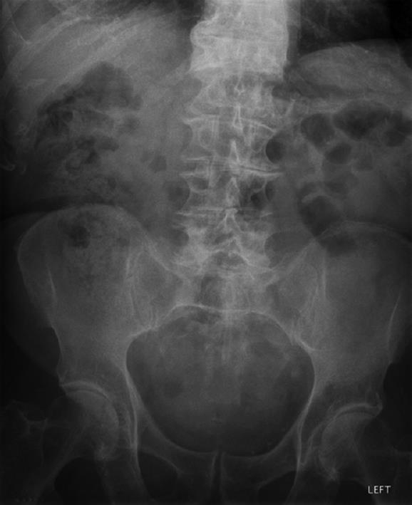
On examination, he was apyrexial with a pulse of 90 bpm. Respiratory rate, oxygen saturations and blood pressure were normal. The abdomen was soft and non-tender though mildly distended. On digital rectal examination a soft bulge was felt on the anterior rectal wall. Blood tests on admission demonstrated dehydration (urea 11.1, creatinine 157) with raised inflammatory markers [C reactive protein (CRP) 278]. An abdominal radiograph (Figure 1) demonstrated some faecal loading but was otherwise featureless.
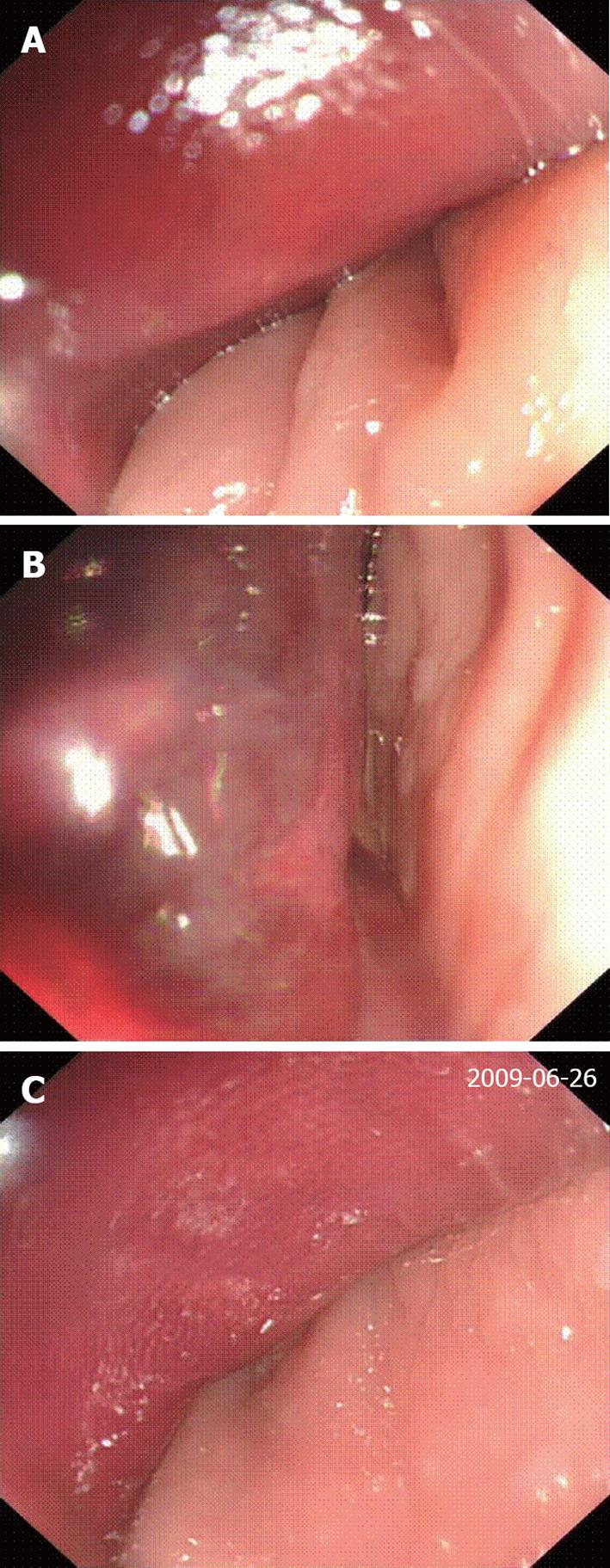
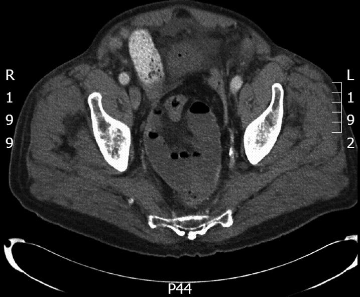
On his second day in the ward the patient became unwell, developing fast atrial fibrillation. He had begun vomiting and a nasogastric tube which was subsequently inserted was draining a high output. A flexible sigmoidoscopy performed because of the rectal mass demonstrated a normal distal rectum. However, 5 cm from the anal verge was a large extra-luminal mass which was purple-pink in colour (Figure 2). This had the appearance of a large haematoma in the rectovesical pouch. No mucosal lesion was identified. A computed tomography scan showed that the pelvis contained fluid-filled small bowel loops, of 3 cm in diameter, clumped together (Figure 3). Prominent small bowel loops were found in the central and upper abdomen with no cause of small bowel obstruction evident.
The patient was taken for a laparotomy which demonstrated distal small bowel obstruction secondary to bowel “incarcerated” within the rectovesical pouch. This small bowel was found to be frankly gangrenous as a result of strangulation, and was tethered to the pelvic brim. He was noted to have a narrow pelvic inlet and underwent resection for the gangrenous small bowel loops. After an uneventful postoperative recovery he was discharged home.
The flexible sigmoidoscopy findings together with the unresolving small bowel obstruction prompted a laparotomy where the diagnosis was confirmed. The palpable rectal mass due to a haematoma in the rectovesical pouch, which was visualised at flexible sigmoidoscopy (Figure 2), was due to the presence of gangrenous small bowel which had become strangulated within the confined area of this patient’s notably narrow pelvic inlet. The rectum, being adjacent to this mass of small bowel, was inflamed and oedematous. This probably led to the formation of a haematoma within the rectal wall, which resulted in this most atypical presenting sign.
In males the rectovesical pouch is formed by the part of the peritoneal cavity between the rectum and bladder. There are instances where disease can localise to this region. The most likely pathology is build-up of fluid and abscesses, for example, secondary to appendicitis[1]. Clinically this may be detected by palpating an anterior rectal wall mass on digital rectal examination.
The differential diagnosis of masses palpable per rectum can be divided into causes that are intrinsic (either intra-luminal or within the rectal wall) or extrinsic. Extrinsic causes put pressure of the rectum resulting in the palpable mass. Rigid/flexible sigmoidoscopy will enable the clinician to determine if the mass is extrinsic as with this patient, where the mucosa appeared uninvolved.
It is believed that the narrow inlet of the pelvic cavity in this patient was responsible for the small bowel becoming trapped. The greater transverse mid-inlet and pelvic outlet diameters in females are anecdotally considered by colorectal surgeons as providing better access in operations deep within the pelvic cavity, making the surgery easier than in males[2,3].
The incarceration and subsequent strangulation of small bowel within the pelvic cavity would be comparable to the pathology involved in internal herniae. In this case the hernia would be represented by the rectovesical pouch with the narrow pelvic inlet behaving as a tight neck. Our case report details the only instance of herniation within the rectovesical pouch of the pelvis.
Strangulation of small bowel within the pelvic cavity is a possibility in males where the inlet may be particularly constricted. This can rapidly lead to ischaemic bowel and therefore significant mortality and morbidity. Awareness of this pathology in patients presenting with atypical small bowel obstruction (SBO) is important as well as recognition that digital rectal examination findings may be indicative of small bowel strangulation.
Peer reviewers: Tang Chung Ngai, MB, BS, Professor, Department of Surgery, Pamela Youde Nethersole Eastern Hospital, 3 Lok Man Road, Chai Wan, Hong Kong, China; Uwe Klinge, MD, Professor, Institute for Applied Medical Engineering AME, Helmholtz Institute, RWTH Aachen Pauwelsstrabe 30, Aachen 52074, Germany
S- Editor Wang JL L- Editor Hughes D E- Editor Lin YP
| 1. | Welborn MB. Postappendiceal abscess in the rectovesical pouch; transrectal drainage. Am J Surg. 1952;83:176-180. |
| 2. | Salerno G, Daniels IR, Brown G, Heald RJ, Moran BJ. Magnetic resonance imaging pelvimetry in 186 patients with rectal cancer confirms an overlap in pelvic size between males and females. Colorectal Dis. 2006;8:772-776. |
| 3. | Li JC, Chu DW, Lee DW, Chan AC. Small-bowel intestinal obstruction caused by an unusual internal hernia. Asian J Surg. 2005;28:62-64. |


