Copyright
©The Author(s) 2023.
World J Gastrointest Surg. May 27, 2023; 15(5): 965-971
Published online May 27, 2023. doi: 10.4240/wjgs.v15.i5.965
Published online May 27, 2023. doi: 10.4240/wjgs.v15.i5.965
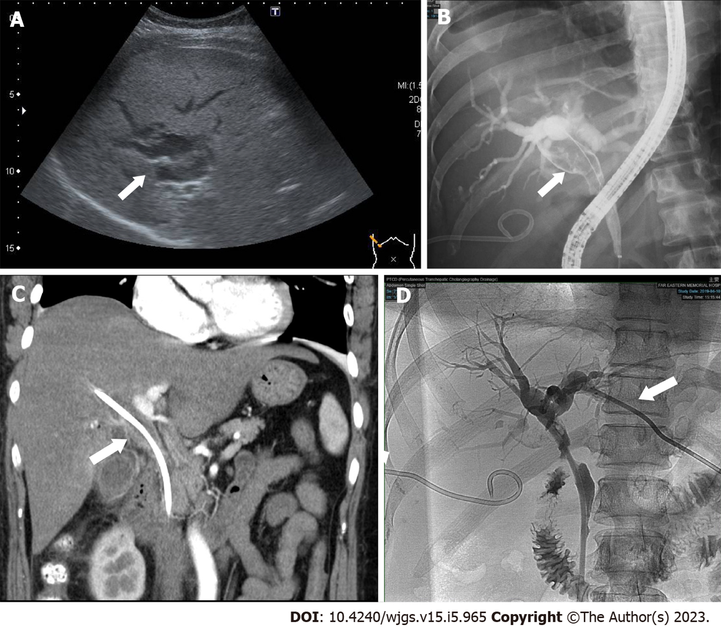
Figure 1 Imaging results.
A: Abdominal sonography showed dilated intrahepatic ducts (arrow); B: The cholangiogram revealed a long filling defect (arrow) from the proximal common bile duct (CBD) to the common hepatic duct; C: Contrast-enhanced computed tomography also showed soft tissue density (arrow) from the proximal CBD to the common hepatic duct; D: Percutaneous transhepatic cholangiography drainage (arrow) was performed on the left intrahepatic duct to alleviate jaundice.
- Citation: Chiang CH, Chen KC, Devereaux B, Chung CS, Kuo KC, Lin CC, Lin CK, Wang HP, Chen KH. Precise mapping of hilar cholangiocarcinoma with a skip lesion by SpyGlass cholangioscopy: A case report. World J Gastrointest Surg 2023; 15(5): 965-971
- URL: https://www.wjgnet.com/1948-9366/full/v15/i5/965.htm
- DOI: https://dx.doi.org/10.4240/wjgs.v15.i5.965


