Published online Dec 27, 2023. doi: 10.4240/wjgs.v15.i12.2932
Peer-review started: August 28, 2023
First decision: November 1, 2023
Revised: November 8, 2023
Accepted: December 8, 2023
Article in press: December 8, 2023
Published online: December 27, 2023
Processing time: 121 Days and 1.7 Hours
Endoscopic submucosal dissection (ESD) can be used for the en-bloc removal of superficial rectal lesions; however, the lack of a traction system makes the procedure long and difficult in the presence of extensive lesions.
A large polyp occupying 2/3 of the rectal circumference and extending 5 cm in length was removed by ESD with the help of laparoscopic forceps introduced via trans-anal rectoscopic assisted minimally invasive surgery, a disposable platform designed to aid in transanal minimally invasive surgery. Traction of the polyp by forceps during the operation was dynamic, and applied at various points and in various directions. The polyp was removed en-bloc without complications in 1 h and 55 min. A sigmoidoscopy performed 50 d later showed normal healing without polyp recurrence.
The technique presented here could overcome the issues caused by lack of traction during ESD for rectal lesions.
Core Tip: Endoscopic submucosal dissection (ESD) can be used for the en-bloc removal of superficial rectal lesions; however, the lack of a traction system makes the procedure long and difficult in the presence of extensive lesions. In this case, the use of trans-anal rectoscopic assisted minimally invasive surgery could overcome the problem of traction during ESD for rectal lesions.
- Citation: Polese L. Removal of a large rectal polyp with endoscopic submucosal dissection-trans-anal rectoscopic assisted minimally invasive surgery hybrid technique: A case report. World J Gastrointest Surg 2023; 15(12): 2932-2937
- URL: https://www.wjgnet.com/1948-9366/full/v15/i12/2932.htm
- DOI: https://dx.doi.org/10.4240/wjgs.v15.i12.2932
Anterior rectal resection with total mesorectal excision (TME), generally performed after neoadjuvant chemioradiotherapy, is a radical treatment for rectal tumours. This treatment modality is highly invasive, and is associated with numerous complications, including high rates (up to 40%) of genitourinary and sexual dysfunction, long term functional bowel impairment and risk of enterostomy[1].
To minimize trauma, early rectal tumours can be treated using approaches other than colorectal resection, that offer better functional results and reduce the risk of an intestinal stoma.
In the 1980s, Buess[2] first described the transanal endoscopic microsurgery (TEM), a minimally invasive surgical technique for resection of small rectal cancers and benign lesions of the low, middle, and upper rectum. This local resection technique can be curative for lesions limited to the organ, without risk of lymph-node metastases.
When performed with oncological radicality, TEM offers significant advantages in terms of morbidity, mortality and functional outcome compared to radical resection. Endoscopic submucosal dissection (ESD) is another interventional procedure suitable for en-bloc resection of early gastrointestinal lesions. ESD is a flexible endoscopic procedure, with reduced risk of recurrence with respect to standard endoscopic mucosal resection, in particular for lesions greater than 20 mm in size.
Although this technique was initially used for the upper gastrointestinal tract, ESD indications have in recent years been extended to include colorectal lesions[3]. In the presence of large polyps, and in particular in those with nodules, ESD should be preferred over EMR due to the lower risk of invasion. Thus, for benign lesions and early cancers located in the rectum both TEM and ESD represent a less invasive alternative to rectal resection.
A previous systematic review[4] compared TEM with ESD performed for large nonpedunculated rectal lesions, preoperatively assessed as non-invasive, and found similar results in term of postoperative complications (8%-8.4%), but higher R0 resection rate for TEM (88.5% vs 74.6%).
Thus full-thickness TEM offers higher rate of R0 resection and low rates of local recurrence; however, opening of rectal wall is associated with an increased risk of peritoneal entry and trauma to the mesorectum, leading to adverse events, including peritonitis. Additionally, according to some researchers, scarring in the mesorectum makes salvage TME much more challenging in cases of unfavourable histological features or recurrence[5].
ESD preserves the deeper layers of the rectal wall and leaves the mesorectum intact. This is particularly advantageous when a large polyp has to be removed, as it can avoid causing severe colorectal strictures.
Some surgeons perform an ESD using the TEM platform and TEM instruments with the aid of hydro-dissection to remove only the superficial plan through a submucosal dissection, with the aim of reducing the deep trauma generally associated with full TEM[6]. From a technical point the theoretical advantage of TEM vs endoscopy is that both hands can be used: this allows the polyp to be grasped and put under tension, allowing better exposure during dissection.
On the other hand, flexible endoscopy is associated with greater freedom of movement, and allows the surgeon to see and treat lesions from different points, even in the presence of large polyps.
Herein, we present an innovative technique in which ESD of a rectal polyp was performed with a flexible endoscope, with the aid of laparoscopic forceps introduced via the trans-anal rectoscopic assisted minimally invasive surgery (ARAMIS) platform.
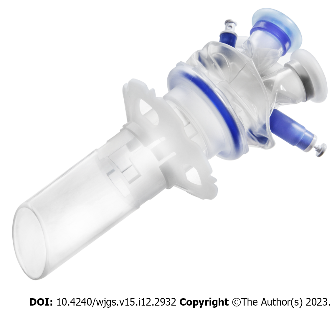
ARAMIS (SapiMed, Alessandria, Italy) is a disposable platform for trans-anal rectoscopic-assisted minimally invasive surgery comprising an all-in-one solution combining a single port access and a rigid rectoscope[7].
The rectoscope has a directional 45° flute opening and an insertion depth that varies between 5-7 and 9-11 cm from the anal verge. The rectoscope is self-sustaining by means of a plastic fixing ring sutured to the perianal skin, and is connected to a disposable, flexible, single access surgery port (Gloveport, Nelis, Bucheon City, South Korea) that has four working channels: Three for instruments up to 5 mm in diameter, and one for instruments up to 12 mm in diameter (Figure 1).
Herein, we present the case of a 76-year-old woman with a large rectal polyp (Video 1). Informed consent was obtained from the patient for publication of this case report.
The polyp was found during a colonoscopy performed for rectal bleeding.
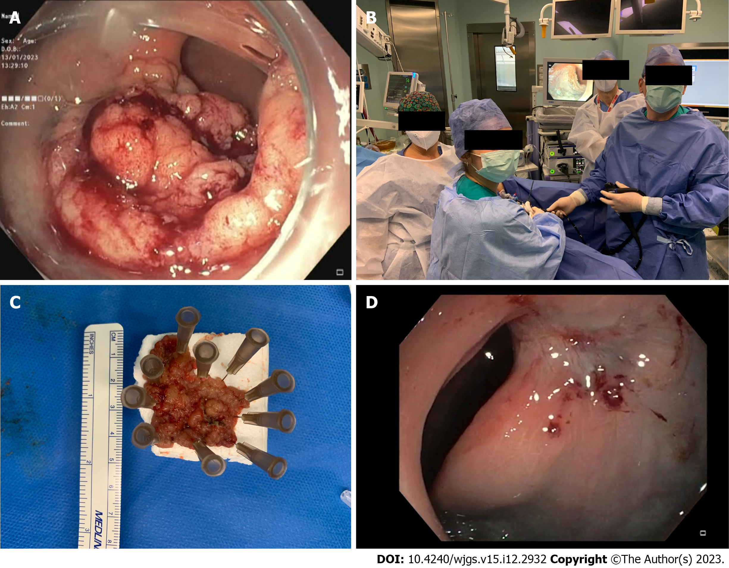
The patient presented with a bleeding rectal polyp. The polyp occupied up to 2/3 of the circumference of the rectum (Figure 2A), extended about 5 cm in length, and was situated from 10 to 15 cm from the anal verge. The lesion was mostly located in the anterior rectal wall. The polyp was found during a colonoscopy performed for bleeding. Preoperative biopsy documented an adenoma with low-grade dysplasia. After informed consent, surgery was performed with the patient in the prone Jackknife position and intubated. The operating team comprised two surgeons and two nurses (Figure 2B).
Biohumoral tests revealed a mild anemia.
At colonoscopy the polyp occupied up to 2/3 of the circumference of the rectum (Figure 2A), extended about 5 cm in length, and was situated from 10 to 15 cm from the anal verge. The lesion was mostly located in the anterior rectal wall.
Preoperative biopsy documented an adenoma with low-grade dysplasia.
After informed consent, surgery was performed with the patient in the prone Jackknife position and intubated. The operating team comprised two surgeons and two nurses (Figure 2B).
During surgery, the ARAMIS was first positioned and secured to the perineum by 2 perianal skin stitches. After port insertion, a standard gastroscope GIFH190 (Olympus, Tokyo, Japan) (external diameter 9.2 mm) with transparent cap, was introduced via the 12 mm working channel, and a laparoscopic dissector was introduced via a 5 mm working channel. We used a laparoscopic dissector instead of a laparoscopic Johan’s grasping forceps because the former has an angulated tip that makes movements in small spaces easier. CO2 insufflation was applied via the endoscope, which was connected to a CO2 insufflator (CO2 Endo Stratus, Medivators, Minneapolis, MN, United States).
At the beginning of surgery, the mucosa was lifted by an endoscopic injection needle, using saline solution with methylene blue, after which submucosal dissection was performed with Dual knife J (Olympus, Tokyo, Japan), which has a channel for water injection during ESD procedure. Submucosal vessels were treated by diathermy using dual knife or Coagrasper Hemostatic Forceps (Olympus, Tokyo, Japan). The ESD procedure was assisted by traction with the dissector, lifting the polyp from different points and in different directions during the various phases of the operation.
The procedure lasted 1 h and 55 min and the polyp was successfully removed en-bloc (Figure 2C). The operation was performed without complications. Final histological examination confirmed the diagnosis of adenoma with low grade dysplasia.
The patient received oral feeding the same day of the operation and was discharged from the hospital on the third postoperative day with a prescription for oral Vaseline oil. A sigmoidoscopy performed 50 d later showed a regular healing without polyp recurrence (Figure 2D).
Insufficient countertraction and poor view of the submucosa makes ESD a difficult and lengthy procedure. Several traction methods have been reported in literature, but traction is usually limited to one direction[8]. One potential solution is pocket creation assisted ESD[9]. This technique is derived from peroral endoscopic myotomy tunnel creation technique, but requires considerable technical skill, especially in the colon. Magnetic anchor-guided ESD has also been proposed as a solution, but requires special devices which are not readily available[10]. Rectal ESD and TEM are the most popular techniques for early rectal neoplasms resection. ESD spares the deep tissue and can be applied to more extensive superficial lesions, avoiding deep rectal openings and strictures. TEM also has the advantage of applying counter-traction during manoeuvres.
The technique presented herein combines the advantages of the two methods, including the close view of the endoscope, which allows easy recognition of even small vessels and easy identification of the submucosal layer. Further, hydro-dissection is simpler and can be performed simultaneously with diathermic dissection, making the procedure simpler than submucosal dissection performed with the TEM technique. Dissection is also facilitated by dedicated endoscopic accessories, which combine diathermy and hydrodissection, such as the hybrid knife (ERBE) or the dual knife J (Olympus), as in the case presented here. Coagulation of the submucosal vessels is carried out with particular accuracy, avoiding thermal damage in depth, with risk of perforation. This is particularly important when the dissection is extended above the peritoneal reflection, where a deep wall opening could cause peritonitis. Submucosal dissection is simultaneously made easier and faster by the traction performed with laparoscopic forceps, introduced through the ARAMIS platform.
The traction performed in this manner is dynamic; it can be applied at various points and in various directions during the operation. In particular, traction is not only performed upwards, but also laterally, cranially or caudally, allowing exposure of different areas of the polyp and the submucosal layer.
Furthermore, the ARAMIS platform can allow the possible insertion of other laparoscopic instruments, for example a needle holder to perform a suture in case of need.
The adjustable rectoscope protects the rectum during manoeuvres with laparoscopic instruments, avoiding the risk of damage. It also keeps the rectum open, when the wall collapses or pressure drops.
In conclusion, ESD-ARAMIS is a new method which can be applied to superficial rectal neoplasms in which ESD is facilitated by the use of laparoscopic instrumentation introduced via the ARAMIS platform. This technique can make the procedure faster and safer, particularly in cases where the removal of large lesions is required.
Provenance and peer review: Unsolicited article; Externally peer reviewed.
Peer-review model: Single blind
Specialty type: Surgery
Country/Territory of origin: Italy
Peer-review report’s scientific quality classification
Grade A (Excellent): A
Grade B (Very good): B
Grade C (Good): C
Grade D (Fair): 0
Grade E (Poor): 0
P-Reviewer: Cai KL, China; Luo W, China; Wang LH, China S-Editor: Yan JP L-Editor: A P-Editor: Yan JP
| 1. | Morino M, Arezzo A, Allaix ME. Transanal endoscopic microsurgery. Tech Coloproctol. 2013;17 Suppl 1:S55-S61. [RCA] [PubMed] [DOI] [Full Text] [Cited by in Crossref: 27] [Cited by in RCA: 20] [Article Influence: 1.7] [Reference Citation Analysis (0)] |
| 2. | Buess G. Endoskopie—von der Diagnostik bis zur neuen Chirurgie. Deutscher Arzte-Verlag, Köln. 1990. |
| 3. | Fuccio L, Ponchon T. Colorectal endoscopic submucosal dissection (ESD). Best Pract Res Clin Gastroenterol. 2017;31:473-480. [RCA] [PubMed] [DOI] [Full Text] [Cited by in Crossref: 20] [Cited by in RCA: 20] [Article Influence: 2.5] [Reference Citation Analysis (0)] |
| 4. | Arezzo A, Passera R, Saito Y, Sakamoto T, Kobayashi N, Sakamoto N, Yoshida N, Naito Y, Fujishiro M, Niimi K, Ohya T, Ohata K, Okamura S, Iizuka S, Takeuchi Y, Uedo N, Fusaroli P, Bonino MA, Verra M, Morino M. Systematic review and meta-analysis of endoscopic submucosal dissection versus transanal endoscopic microsurgery for large noninvasive rectal lesions. Surg Endosc. 2014;28:427-438. [RCA] [PubMed] [DOI] [Full Text] [Cited by in Crossref: 120] [Cited by in RCA: 103] [Article Influence: 9.4] [Reference Citation Analysis (0)] |
| 5. | Guerrieri M, Gesuita R, Ghiselli R, Lezoche G, Budassi A, Baldarelli M. Treatment of rectal cancer by transanal endoscopic microsurgery: experience with 425 patients. World J Gastroenterol. 2014;20:9556-9563. [RCA] [PubMed] [DOI] [Full Text] [Full Text (PDF)] [Cited by in CrossRef: 41] [Cited by in RCA: 48] [Article Influence: 4.4] [Reference Citation Analysis (0)] |
| 6. | Kouladouros K, Baral J. Transanal endoscopic microsurgical submucosal dissection (TEM-ESD): A novel approach to the local treatment of early rectal cancer. Surg Oncol. 2021;39:101662. [RCA] [PubMed] [DOI] [Full Text] [Cited by in Crossref: 7] [Cited by in RCA: 3] [Article Influence: 0.8] [Reference Citation Analysis (0)] |
| 7. | Polese L, Rizzato R, Porzionato A, Da Dalt G, Bressan A, De Caro R, Merigliano S. An evaluation of trans-anal rectoscopic-assisted minimally invasive surgery (ARAMIS): a new platform for transanal surgery. Int J Colorectal Dis. 2020;35:1681-1687. [RCA] [PubMed] [DOI] [Full Text] [Full Text (PDF)] [Cited by in Crossref: 2] [Cited by in RCA: 2] [Article Influence: 0.4] [Reference Citation Analysis (0)] |
| 8. | Yamasaki Y, Takeuchi Y, Uedo N, Kanesaka T, Kato M, Hamada K, Tonai Y, Matsuura N, Akasaka T, Hanaoka N, Higashino K, Ishihara R, Okada H, Iishi H. Efficacy of traction-assisted colorectal endoscopic submucosal dissection using a clip-and-thread technique: A prospective randomized study. Dig Endosc. 2018;30:467-476. [RCA] [PubMed] [DOI] [Full Text] [Cited by in Crossref: 63] [Cited by in RCA: 92] [Article Influence: 13.1] [Reference Citation Analysis (0)] |
| 9. | Pei Q, Qiao H, Zhang M, Wang G, Feng H, Pan J, Shi Y. Pocket-creation method versus conventional method of endoscopic submucosal dissection for superficial colorectal neoplasms: a meta-analysis. Gastrointest Endosc. 2021;93:1038-1046.e4. [RCA] [PubMed] [DOI] [Full Text] [Cited by in Crossref: 10] [Cited by in RCA: 24] [Article Influence: 6.0] [Reference Citation Analysis (0)] |
| 10. | Matsuzaki I, Hattori M, Yamauchi H, Goto N, Iwata Y, Yokoi T, Tsunemi M, Kobayashi M, Yamamura T, Miyahara R. Magnetic anchor-guided endoscopic submucosal dissection for colorectal tumors (with video). Surg Endosc. 2020;34:1012-1018. [RCA] [PubMed] [DOI] [Full Text] [Cited by in Crossref: 12] [Cited by in RCA: 23] [Article Influence: 3.8] [Reference Citation Analysis (0)] |


