Copyright
©The Author(s) 2015.
World J Gastrointest Surg. Dec 27, 2015; 7(12): 398-402
Published online Dec 27, 2015. doi: 10.4240/wjgs.v7.i12.398
Published online Dec 27, 2015. doi: 10.4240/wjgs.v7.i12.398
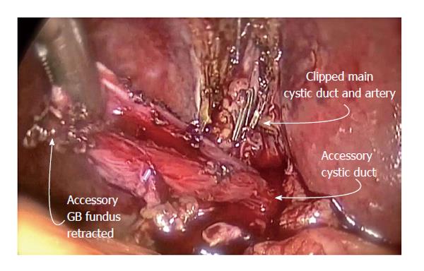
Figure 1 The accessory gallbladder shown here is dissected of the shared liver bed with the main gallbladder.
Clips are placed on the divided main cystic duct and artery. GB: Gallbladder.
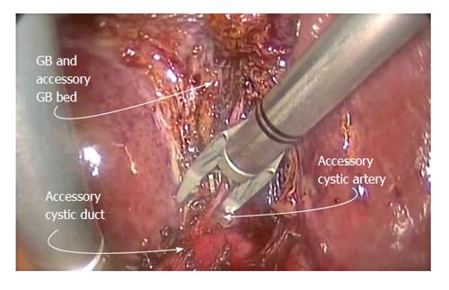
Figure 2 Further dissection of the accessory gallbladder revealed the accessory cystic artery, which helped in the identification of the cystic structure as an accessory gallbladder.
The accessory cystic artery is dissected off the accessory cystic duct situated below, clipped and divided. A cholangiogram through the accessory cystic duct was performed. GB: Gallbladder.
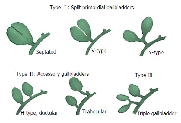
Figure 3 Harlaftis’s classification of anatomical variations of accessory gallbladders.
- Citation: Cozacov Y, Subhas G, Jacobs M, Parikh J. Total laparoscopic removal of accessory gallbladder: A case report and review of literature. World J Gastrointest Surg 2015; 7(12): 398-402
- URL: https://www.wjgnet.com/1948-9366/full/v7/i12/398.htm
- DOI: https://dx.doi.org/10.4240/wjgs.v7.i12.398


