Copyright
©2013 Baishideng Publishing Group Co.
World J Gastrointest Surg. Jul 27, 2013; 5(7): 224-228
Published online Jul 27, 2013. doi: 10.4240/wjgs.v5.i7.224
Published online Jul 27, 2013. doi: 10.4240/wjgs.v5.i7.224
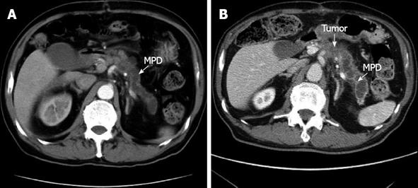
Figure 1 Computed tomography scan demonstrating the dilatation of the main pancreatic duct (A) and a tumor of the pancreatic body which invaded the celiac artery after the abdominal aortic aneurysm operation (B).
MPD: Main pancreatic duct.
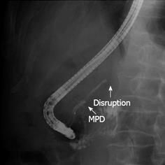
Figure 2 Endoscopic retrograde cholangiopancreatography showing a stricture of the main pancreatic duct in the body of the pancreas.
MPD: Main pancreatic duct.
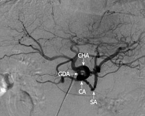
Figure 3 Abdominal angiography showing splenic and celiac arteries involved with a solid tumor in the pancreatic body and the gastroduodenal artery arising from the area close to the celiac.
SA: Splenic CA: Celiac axis; CHA: Common hepatic artery; GDA: Gastroduodenal artery.
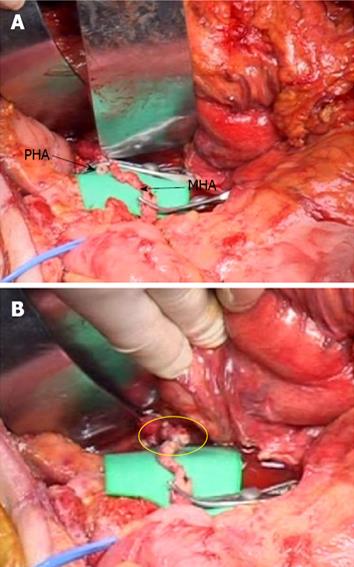
Figure 4 Operative photograph showing that reconstruction was completed.
End-to-end anastomosis was performed between the proper hepatic artery (PHA) and the middle colic artery (MCA) (yellow circle). A: Before anastomosis; B: After anastomosis.
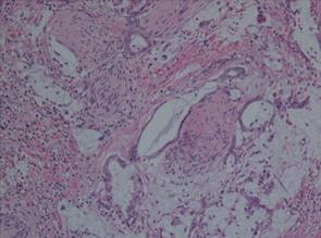
Figure 5 In the pathological findings, there was a prominent formation of mucinous nodules and mucinous carcinoma including large quantities of mucus.
Final histolopathological diagnosis of the resected specimen shows mucinous carcinoma (with an intraductal papillary-mucinous tumor, Hematoxylin eosin staining, ×100).
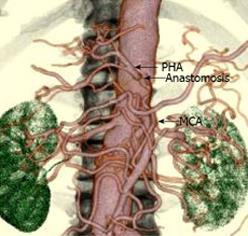
Figure 6 Postoperative evaluation of the proper hepatic artery by 3D-computer tomography angiography.
The arrows indicate the anastomosis between the proper hepatic artery (PHA) and the middle colic artery (MCA).
- Citation: Suzuki H, Hosouchi Y, Sasaki S, Araki K, Kubo N, Watanabe A, Kuwano H. Reconstruction of the hepatic artery with the middle colic artery is feasible in distal pancreatectomy with celiac axis resection: A case report. World J Gastrointest Surg 2013; 5(7): 224-228
- URL: https://www.wjgnet.com/1948-9366/full/v5/i7/224.htm
- DOI: https://dx.doi.org/10.4240/wjgs.v5.i7.224


