Copyright
©2013 Baishideng Publishing Group Co.
World J Gastrointest Surg. Jun 27, 2013; 5(6): 210-215
Published online Jun 27, 2013. doi: 10.4240/wjgs.v5.i6.210
Published online Jun 27, 2013. doi: 10.4240/wjgs.v5.i6.210
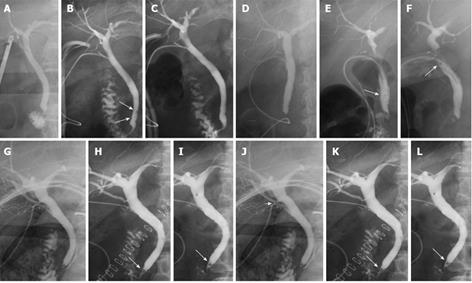
Figure 1 Cholangiography performed in patients.
A: Intraoperative cholangiography (IOC) could not identify a filling defect; B: On postoperative day (POD) 7, cholangiography demonstrated a 2 mm-diameter stone (white arrow); C: Topical glyceryl trinitrate (GTN) infusion using cystic duct tube (C-tube) was performed (on the following day the retained stone disappeared) (white arrow); D: IOC could not identify a filling defect; E: On POD 9, cholangiography showed 2 mm-diameter stones at distal common bile duct (CBD) (white arrow); F: GTN was infused three times and the retained stones were removed (white arrow); G: IOC could not show a filling defect, but contrast media did not flow into duodenum; H: Cholangiography showed a 3 mm-diameter floating stone on POD 5 (white arrow); I: Isosorbide dinitrate was infused 6 times, but the stone was not removed (white arrow); J: Since an injury to posterior superior bile duct was suspected by IOC (white arrow head); K: C-tube was placed. On POD 7, cholangiography showed a 6 mm-diameter retained stone at distal CBD without biliary leak (white arrow); L: GTN was infused 4 times, but the stone could not be removed (white arrow).
- Citation: Shoji M, Sakuma H, Yoshimitsu Y, Maeda T, Nakai M, Ueda H. Topical nitrate drip infusion using cystic duct tube for retained bile duct stone: A six patients case series. World J Gastrointest Surg 2013; 5(6): 210-215
- URL: https://www.wjgnet.com/1948-9366/full/v5/i6/210.htm
- DOI: https://dx.doi.org/10.4240/wjgs.v5.i6.210


