Copyright
©2012 Baishideng Publishing Group Co.
World J Gastrointest Surg. Oct 27, 2012; 4(10): 238-245
Published online Oct 27, 2012. doi: 10.4240/wjgs.v4.i10.238
Published online Oct 27, 2012. doi: 10.4240/wjgs.v4.i10.238
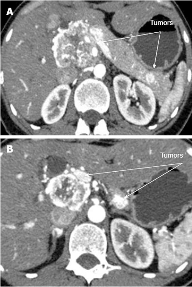
Figure 1 Computed tomography arterial phase.
А: Hypervascular tumors of pancreatic head and tail; В: Hypervascular tumors of pancreatic head and body.
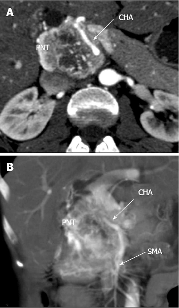
Figure 2 Computed tomography arterial phase.
А: Axial view. Common hepatic artery (CHA) passing through pancreatic parenchyma affected by tumor; В: Coronal view. Hepatomesenteric trunk dividing into superior mesenteric artery and CHA. CHA running within pancreatic tissue (transpancreatic course). SMA: Superior mesenteric artery; PNT: Pancreatic neuroendocrine tumor.
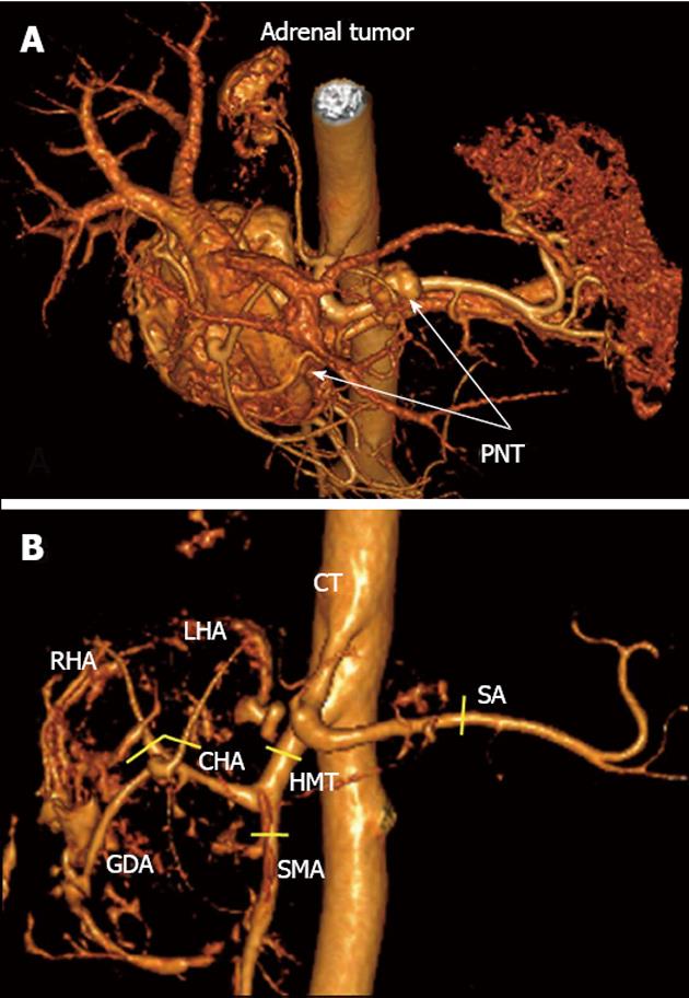
Figure 3 Three-dimensional computed tomography angiography after omission of renal artery image.
А: Hypervascular tumors of pancreatic head and body [pancreatic neuroendocrine tumors (PNTs)], hypervascular right adrenal tumor (tail tumor not shown), right adrenal artery originating from celiac trunk (CT); B: Celiaco-mesenterial arterial architecture found, common hepatic artery and superior mesenteric artery arising from hepatomesenteric trunk (Michels, type IX). GDA: Gastroduodenal artery; LHA: Left hepatic artery; RHA: Right hepatic artery; SMA: Superior mesenteric artery; CHA: Common hepatic artery; SA: Splenic artery; HMT: Hepato mesenteric trunk. Yellow segments indicate lines of arterial resections.
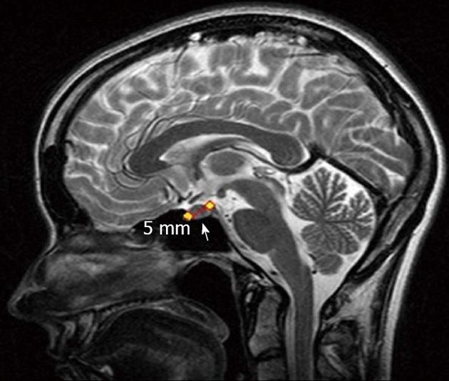
Figure 4 Magnetic resonance imaging.
Pituitary adenoma (arrow).
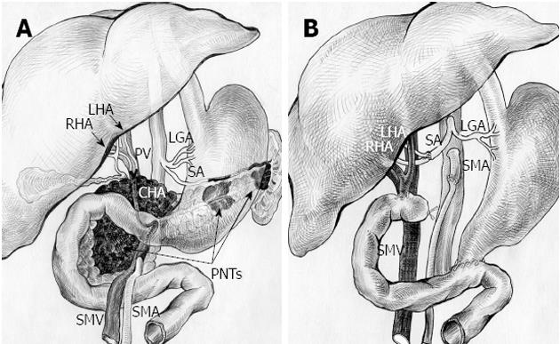
Figure 5 Schematic sketch of procedure with omission of right adrenal tumor.
А: Before surgery. Tumors of pancreatic head, body and tail (pancreatic neuroendocrine tumors, PNTs). Pancreatic head tumor involving site of hepatomesenteric trunk division into superior mesenteric artery (SMA) and common hepatic artery (CHA) together with all of CHA, proper hepatic artery (PHA), gastroduodenal artery (GDA) (omitted) and right and left hepatic arteries (RHA and LHA) bifurcation; B: After total duodenopancreatectomy, splenectomy with SMA, LHA and RHA resection, excision of CHA and PHA and right adrenalectomy. SMA is sutured to the root of resected hepatomesenteric trunk. Splenic artery (SA) resected, U-turned and sutured to newly reconstructed LHA and RHA union. LGA: Left gastric artery; PV: Portal vein; SMV: Superior mesenteric vein.
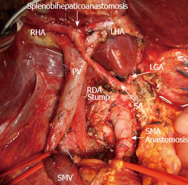
Figure 6 View of operating field after total duodenopancreatectomy and splenectomy with superior mesenteric artery, left hepatic artery and right hepatic resection, excision of common hepatic artery and proper hepatic artery and completion of vascular reconstruction prior to right adrenalectomy.
Superior mesenteric artery (SMA) sutured to the root of resected hepatomesenteric trunk (SMA anastomosis). Splenic artery (SA) resected, U-turned and sutured to newly remade left hepatic artery (LHA) and right hepatic (RHA) junction (splenobihepaticoanastomosis). LGA: Left gastric artery; SMV: Superior mesenteric vein; PV: Portal vein; RDA: Right (inferior) diaphragmatic artery.
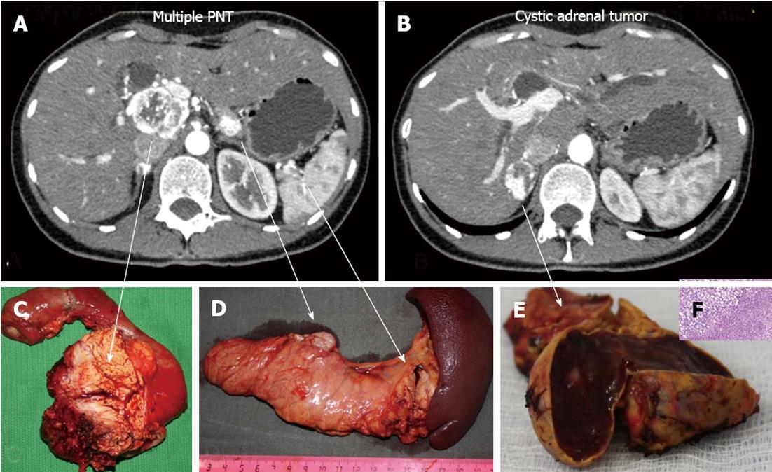
Figure 7 Histopathology: pancreatic tumors (C, D) and right adrenal cystic tumor (E, F) fully compatible with their computed tomography-predicted locations (A, B).
F: Microscopy; HE stain × 200. Right adrenocortical clear cell adenoma.
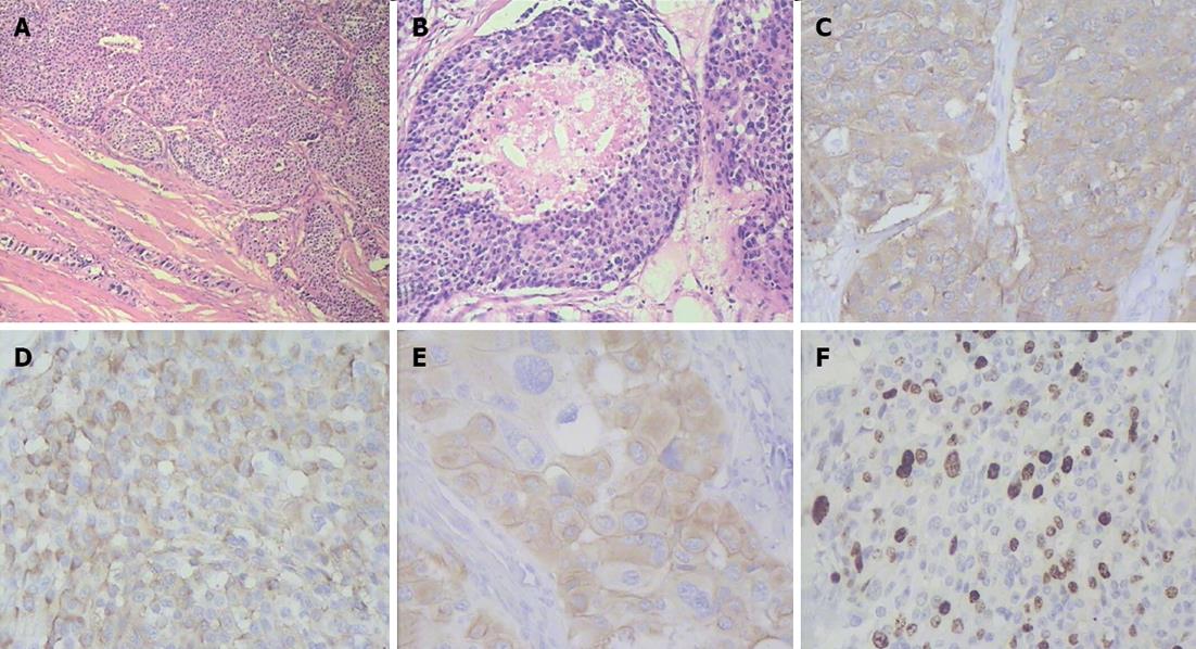
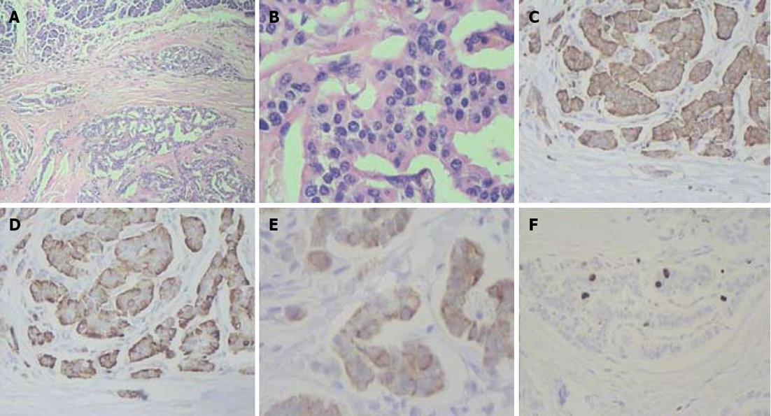
Figure 8 Histopathogical features and immunohistochemical characteristics of pancreatic head neuroendocrine carcinoma.
A: Predominantly solid type of tumor cell growth, HE stain, × 75; B: Comedo-type structure with necrotic focus, HE stain, × 150; C: Strong synaptophysin expression in tumor cells, × 200; D: Strong diffuse chromogranin A expression in tumor cells, × 200; E: Cytokeratin 19 expression in tumor cells, × 200; F: A Ki-67 proliferative index of 40% by immunohistochemistry, × 200.
Figure 9 Histlopathological and immunohistochemical patterns of pancreatic body and tail neuroendocrine tumors.
A: Organoid appearance of tumor cell growth, HE stain, × 75; B: Typical trabecular architecture formed by homogenous small cells, HE stain, × 400; C: Synaptophysin expression in tumor cells, × 200; D: Tumor cells positive for chromogranin A, × 200; E: Tumor cells positive for cytokeratin 19, × 200; F: A Ki-67 proliferative index of 3% by immunohistochemistry, × 200.
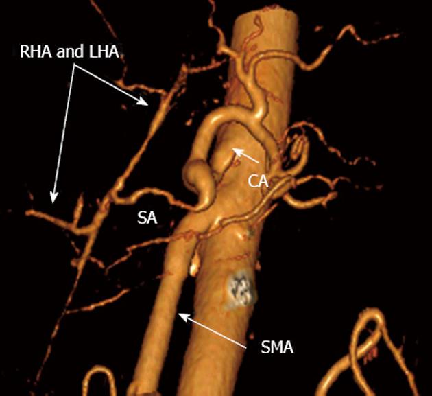
Figure 10 Three-dimensional computed tomography angiograpy (after renal artery image removal) 15 mo post-surgery.
Patent nonstenotic superior mesenteric artery (SMA) and arterial splenobihepaticoanastomosis providing ample adequate blood supply to liver. SА: Splenic; LHA: Left hepatic; RHA: Right hepatic; CA: Celiac artery.
- Citation: Egorov VI, Kharazov AF, Pavlovskaya AI, Petrov RV, Starostina NS, Kondratiev EV, Filippova EM. Extensive multiarterial resection attending total duodenopancreatectomy and adrenalectomy for MEN-1-associated neuroendocrine carcinomas. World J Gastrointest Surg 2012; 4(10): 238-245
- URL: https://www.wjgnet.com/1948-9366/full/v4/i10/238.htm
- DOI: https://dx.doi.org/10.4240/wjgs.v4.i10.238


