Copyright
©2012 Baishideng Publishing Group Co.
World J Gastrointest Surg. Oct 27, 2012; 4(10): 238-245
Published online Oct 27, 2012. doi: 10.4240/wjgs.v4.i10.238
Published online Oct 27, 2012. doi: 10.4240/wjgs.v4.i10.238
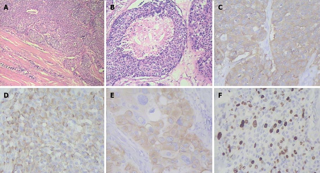
Figure 8 Histopathogical features and immunohistochemical characteristics of pancreatic head neuroendocrine carcinoma.
A: Predominantly solid type of tumor cell growth, HE stain, × 75; B: Comedo-type structure with necrotic focus, HE stain, × 150; C: Strong synaptophysin expression in tumor cells, × 200; D: Strong diffuse chromogranin A expression in tumor cells, × 200; E: Cytokeratin 19 expression in tumor cells, × 200; F: A Ki-67 proliferative index of 40% by immunohistochemistry, × 200.
- Citation: Egorov VI, Kharazov AF, Pavlovskaya AI, Petrov RV, Starostina NS, Kondratiev EV, Filippova EM. Extensive multiarterial resection attending total duodenopancreatectomy and adrenalectomy for MEN-1-associated neuroendocrine carcinomas. World J Gastrointest Surg 2012; 4(10): 238-245
- URL: https://www.wjgnet.com/1948-9366/full/v4/i10/238.htm
- DOI: https://dx.doi.org/10.4240/wjgs.v4.i10.238


