Copyright
©2010 Baishideng Publishing Group Co.
World J Gastrointest Surg. Sep 27, 2010; 2(9): 295-298
Published online Sep 27, 2010. doi: 10.4240/wjgs.v2.i9.295
Published online Sep 27, 2010. doi: 10.4240/wjgs.v2.i9.295
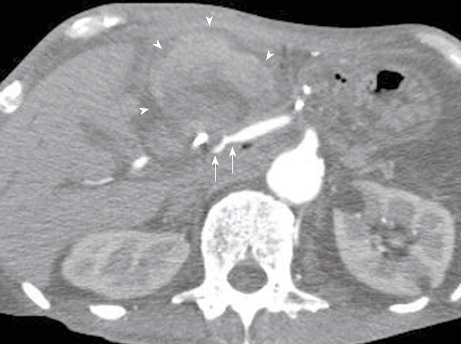
Figure 1 Abdominal computed tomography.
Narrowing of the common hepatic artery and a 7.0 cm × 7.0 cm huge mass with no contrast (arrowheads) extending into the ventral and cranial aspect of the constriction of the common hepatic artery (arrows).
Figure 2 Placement of covered stent graft for pseudoaneurysm of common hepatic artery.
A: Angiography revealed a pseudoaneurysm arising at the common hepatic artery (white arrows) and the dilated lumen of the common hepatic artery (arrowheads); B: A guide wire was advanced past the pseudoaneurysm and a 4 mm x 14 mm covered stent was deployed (arrows); C: Angiography revealed extravasation of contrast medium from the proximal edge of the covered stent (arrow). A dehiscence seemed to occur at the fragile arterial wall; D: An additional covered stent (arrows) was placed partly under lapping the first stent (black arrows).
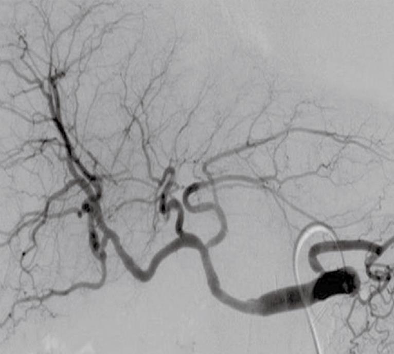
Figure 3 Both hemostasis and blood flow to the liver was confirmed by angiography.
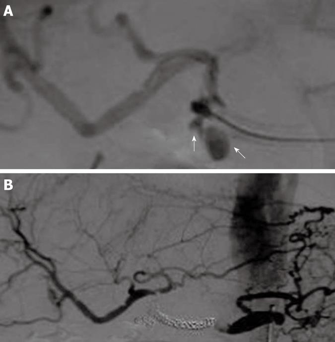
Figure 4 Microcoil embolization in the lumen of the covered stent.
A: Angiography revealed a pseudoaneurysm in the proper hepatic artery at the distal edge of the covered stent (arrows); B: Arteriogram shows the complete exclusion of the common hepatic artery, complete cessation of bleeding and blood flow to the liver via the anastomotic branch of the left gastric artery.
- Citation: Tanaka K, Ohigashi H, Takahashi H, Gotoh K, Yamada T, Miyashiro I, Yano M, Ishikawa O. Successful embolization assisted by covered stents for a pseudoaneurysm following pancreatic surgery. World J Gastrointest Surg 2010; 2(9): 295-298
- URL: https://www.wjgnet.com/1948-9366/full/v2/i9/295.htm
- DOI: https://dx.doi.org/10.4240/wjgs.v2.i9.295


