Copyright
©2010 Baishideng Publishing Group Co.
World J Gastrointest Surg. Sep 27, 2010; 2(9): 295-298
Published online Sep 27, 2010. doi: 10.4240/wjgs.v2.i9.295
Published online Sep 27, 2010. doi: 10.4240/wjgs.v2.i9.295
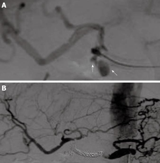
Figure 4 Microcoil embolization in the lumen of the covered stent.
A: Angiography revealed a pseudoaneurysm in the proper hepatic artery at the distal edge of the covered stent (arrows); B: Arteriogram shows the complete exclusion of the common hepatic artery, complete cessation of bleeding and blood flow to the liver via the anastomotic branch of the left gastric artery.
- Citation: Tanaka K, Ohigashi H, Takahashi H, Gotoh K, Yamada T, Miyashiro I, Yano M, Ishikawa O. Successful embolization assisted by covered stents for a pseudoaneurysm following pancreatic surgery. World J Gastrointest Surg 2010; 2(9): 295-298
- URL: https://www.wjgnet.com/1948-9366/full/v2/i9/295.htm
- DOI: https://dx.doi.org/10.4240/wjgs.v2.i9.295


