
















Copyright: ©Author(s) 2026.
World J Gastrointest Surg. Nov 27, 2026; 18(11): 125633
Published online Nov 27, 2026. doi: 10.4240/wjgs.125633
Published online Nov 27, 2026. doi: 10.4240/wjgs.125633
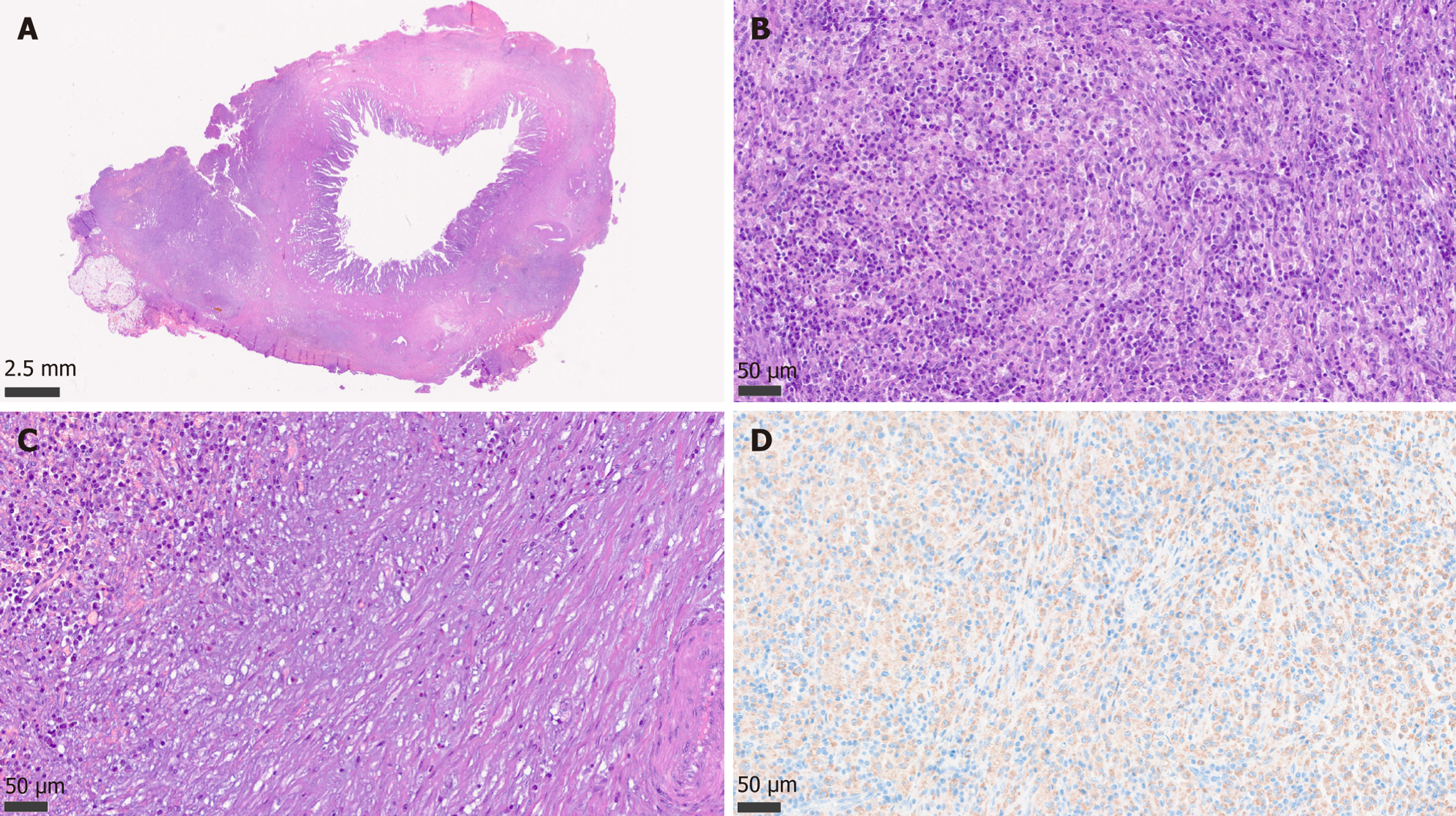
Figure 1 Histologic features of xanthogranulomatous cholecystitis.
A: Low-power view showing an irregularly and unevenly thickened gallbladder wall in a case of xanthogranulomatous cholecystitis; B: The perimuscular soft tissues show a dense inflammatory infiltrate predominantly composed of numerous foamy histiocytes and lymphocytes. No Michaelis-Gutmann bodies, which are characteristic of malakoplakia, are identified; C: In addition to the inflammatory infiltrate, fibroblasts and fibrotic tissue displaying a vaguely storiform pattern are present (right side), potentially posing diagnostic challenges with neoplastic diseases; D: CD68 (PGM1) immunohistochemical staining highlights the predominant histiocytic component.
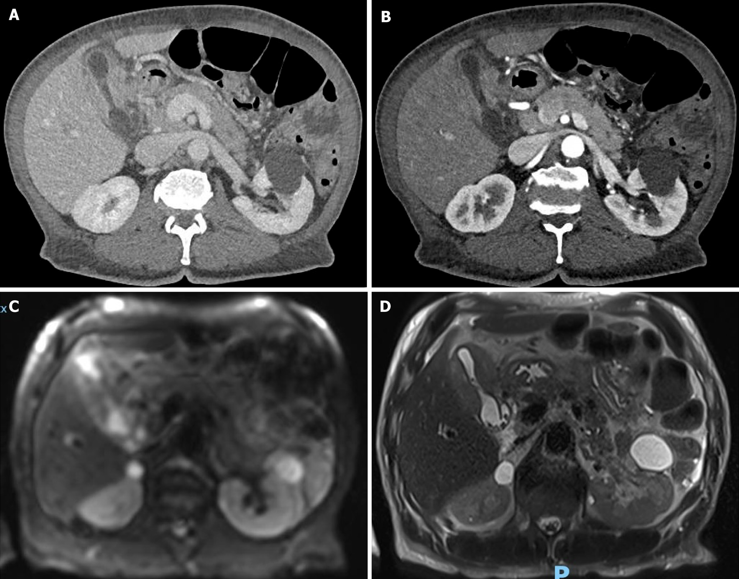
Figure 2 Multimodal preoperative imaging of xanthogranulomatous cholecystitis mimicking gallbladder carcinoma.
A and B: Contrast-enhanced computed tomography in the portal venous and arterial phases showing marked irregular thickening of the gallbladder wall and loss of the cleavage plane with the adjacent liver parenchyma; C: Diffusion-weighted magnetic resonance imaging demonstrating marked diffusion restriction in the gallbladder body-fundus region, further raising suspicion for a neoplastic lesion; D: T2-weighted magnetic resonance imaging showing abnormal gallbladder wall thickening and pericholecystic inflammatory changes, with poor demarcation from the adjacent hepatic parenchyma.
- Citation: Cicerone O, Colletto L, Pellizzari B, Ciuffardi A, Samarawickrama Kodikarage HD, Tushe M, D’Ambrosio G, Vanoli A, Maestri M. Xanthogranulomatous cholecystitis mimicking gallbladder carcinoma. World J Gastrointest Surg 2026; 18(11): 125633
- URL: https://www.wjgnet.com/1948-9366/full/v18/i11/125633.htm
- DOI: https://dx.doi.org/10.4240/wjgs.125633