
















Copyright: ©Author(s) 2026.
World J Gastrointest Surg. Nov 27, 2026; 18(11): 124255
Published online Nov 27, 2026. doi: 10.4240/wjgs.124255
Published online Nov 27, 2026. doi: 10.4240/wjgs.124255
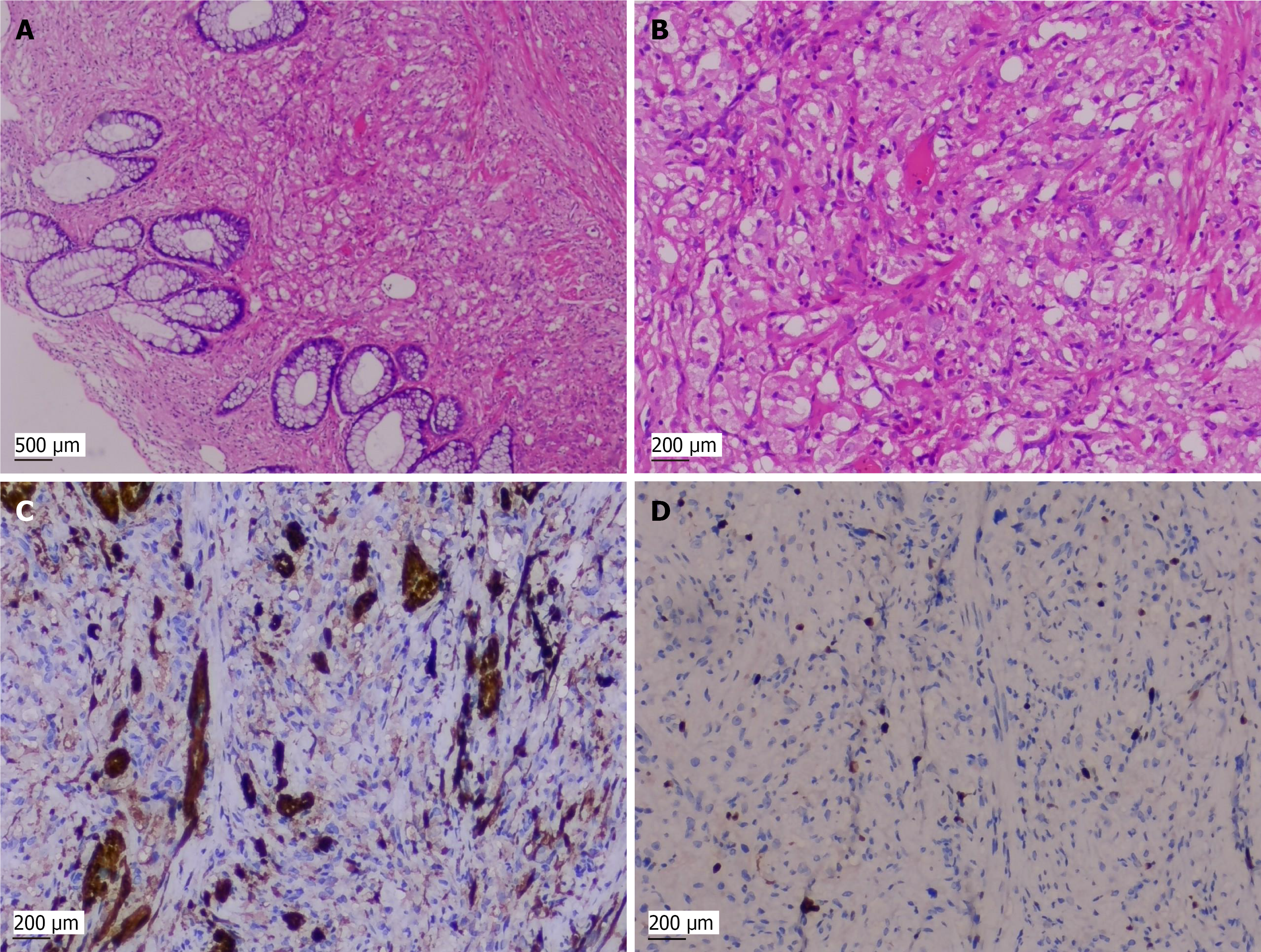
Figure 1 Histopathological and immunohistochemical findings of the rectal granular cell tumor.
A: Tumor cells arranged in nests (× 40); B: Tumor cells with abundant eosinophilic granular cytoplasm (× 100); C: Positive immunohistochemical staining for S-100 protein; D: Ki-67 Labeling index of 3%.
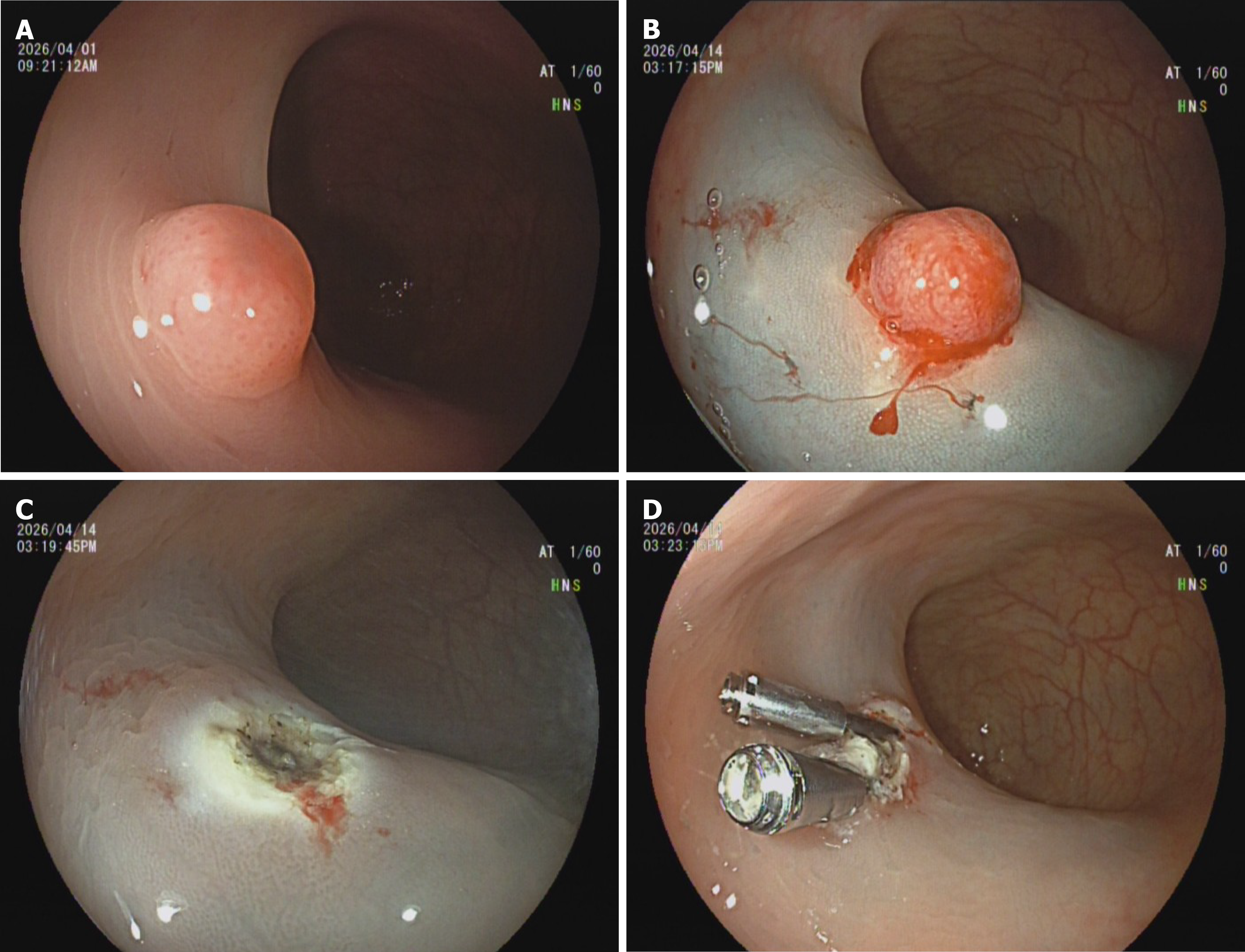
Figure 2 Endoscopic findings and treatment of the rectal lesion.
A: Endoscopic image showing a 6-mm polypoid lesion in the rectum; B: Submucosal injection of methylene blue and glycerol fructose before resection; C: Post-resection wound surface; D: Wound surface with clips.
- Citation: Zhong J, Lu YY, Xiao MY, Wu XQ. Rectal granular cell tumor treated by endoscopic resection: A case report and review of literature. World J Gastrointest Surg 2026; 18(11): 124255
- URL: https://www.wjgnet.com/1948-9366/full/v18/i11/124255.htm
- DOI: https://dx.doi.org/10.4240/wjgs.124255