Copyright
©The Author(s) 2025.
World J Gastrointest Surg. Aug 27, 2025; 17(8): 108963
Published online Aug 27, 2025. doi: 10.4240/wjgs.v17.i8.108963
Published online Aug 27, 2025. doi: 10.4240/wjgs.v17.i8.108963
Figure 1 Macroscopic and imaging examinations of extramammary Paget's disease.
A: Preoperative anal appearance showed atypical perianal eczema-like changes; B: Enteroscopic presentation of anal canal mass, suggesting an infiltrative growth in the 1/2 circle of the posterior wall of the anal canal; C: Preoperative magnetic resonance imaging (as indicated by the white arrow): Suggesting inhomogeneous circumferential thickening of the intestinal wall (about 3/4 circle), downward extension of the anal canal, characterized by the posterior wall, with the posterior wall being thickest by about 1.2 cm, involving the upper and lower diameters of the intestinal canal of about 3.1 cm, and narrowing of the corresponding lumens.
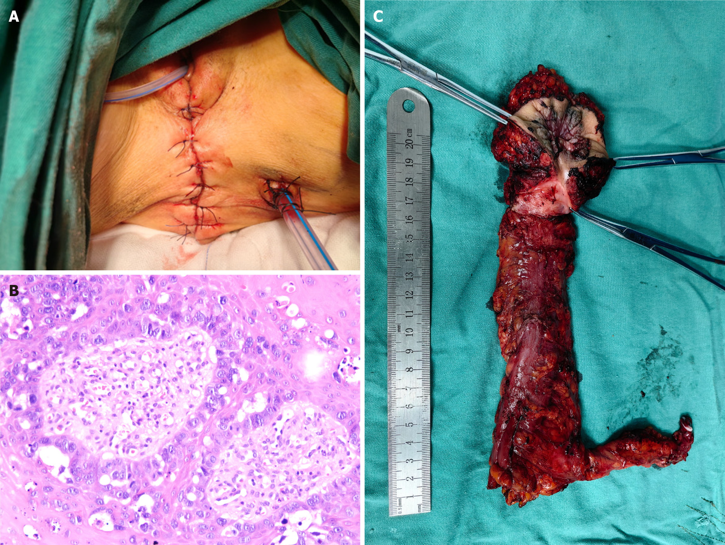
Figure 2 Postoperative appearance, specimen and postoperative immunohistochemistry.
A: Postoperative view of the anal appearance after perineal stage I reconstruction following extended excision of perianal skin; B: Surgical excision specimen, showing rectoanal and perianal extended excised tissues; C: Postoperative immunohistochemistry, Hematoxylin-eosin-stained blood (200 ×) to microscopically visualize the morphology of Paget's cells.
- Citation: Wu SW, Rong Y, Chen GJ, Cao XS, Xie ZY, Wu B, Huang HC, Wang ZW, Wu XX. Anal adenocarcinoma with perianal Paget's disease: A case report. World J Gastrointest Surg 2025; 17(8): 108963
- URL: https://www.wjgnet.com/1948-9366/full/v17/i8/108963.htm
- DOI: https://dx.doi.org/10.4240/wjgs.v17.i8.108963


