Copyright
©The Author(s) 2025.
World J Gastrointest Surg. Aug 27, 2025; 17(8): 108963
Published online Aug 27, 2025. doi: 10.4240/wjgs.v17.i8.108963
Published online Aug 27, 2025. doi: 10.4240/wjgs.v17.i8.108963
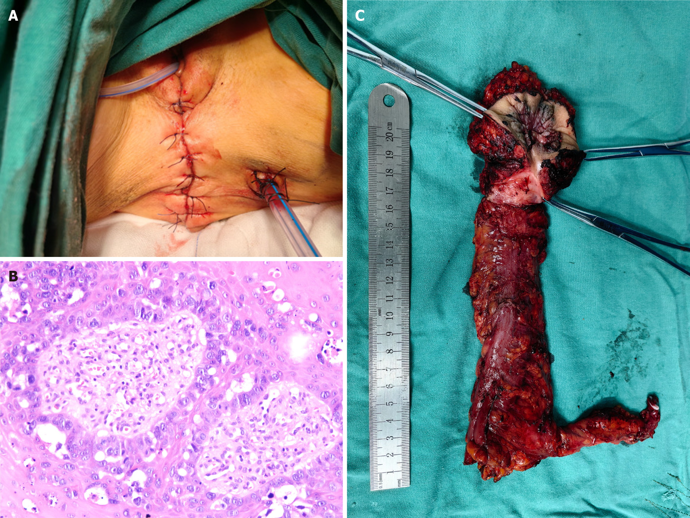
Figure 2 Postoperative appearance, specimen and postoperative immunohistochemistry.
A: Postoperative view of the anal appearance after perineal stage I reconstruction following extended excision of perianal skin; B: Surgical excision specimen, showing rectoanal and perianal extended excised tissues; C: Postoperative immunohistochemistry, Hematoxylin-eosin-stained blood (200 ×) to microscopically visualize the morphology of Paget's cells.
- Citation: Wu SW, Rong Y, Chen GJ, Cao XS, Xie ZY, Wu B, Huang HC, Wang ZW, Wu XX. Anal adenocarcinoma with perianal Paget's disease: A case report. World J Gastrointest Surg 2025; 17(8): 108963
- URL: https://www.wjgnet.com/1948-9366/full/v17/i8/108963.htm
- DOI: https://dx.doi.org/10.4240/wjgs.v17.i8.108963


