Copyright
©The Author(s) 2025.
World J Gastrointest Surg. Jun 27, 2025; 17(6): 107033
Published online Jun 27, 2025. doi: 10.4240/wjgs.v17.i6.107033
Published online Jun 27, 2025. doi: 10.4240/wjgs.v17.i6.107033
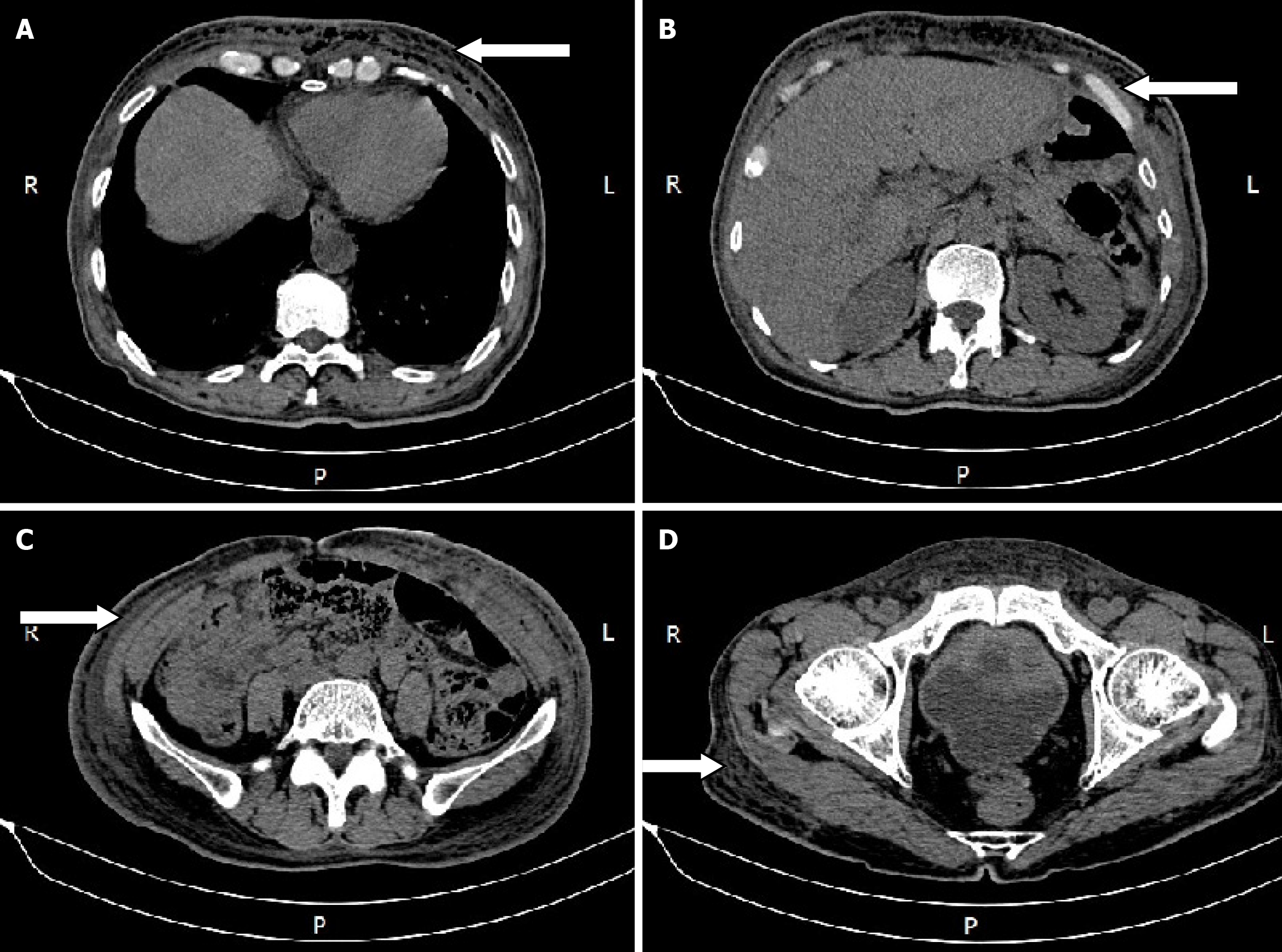
Figure 1 Chest and abdominal wall abscess shown on computed tomography.
A: Abscess on chest wall (arrow: Abscess on chest wall); B-D: Abscess all over abdominal wall (arrow: Abscess all over abdominal wall).
Figure 2 Upper gastrointestinal radiography showed that the gastric wall was irregular and tentacle-like.
A and B: View from front; C: View from behind. Arrow: The gastric wall was irregular and tentacle-like.
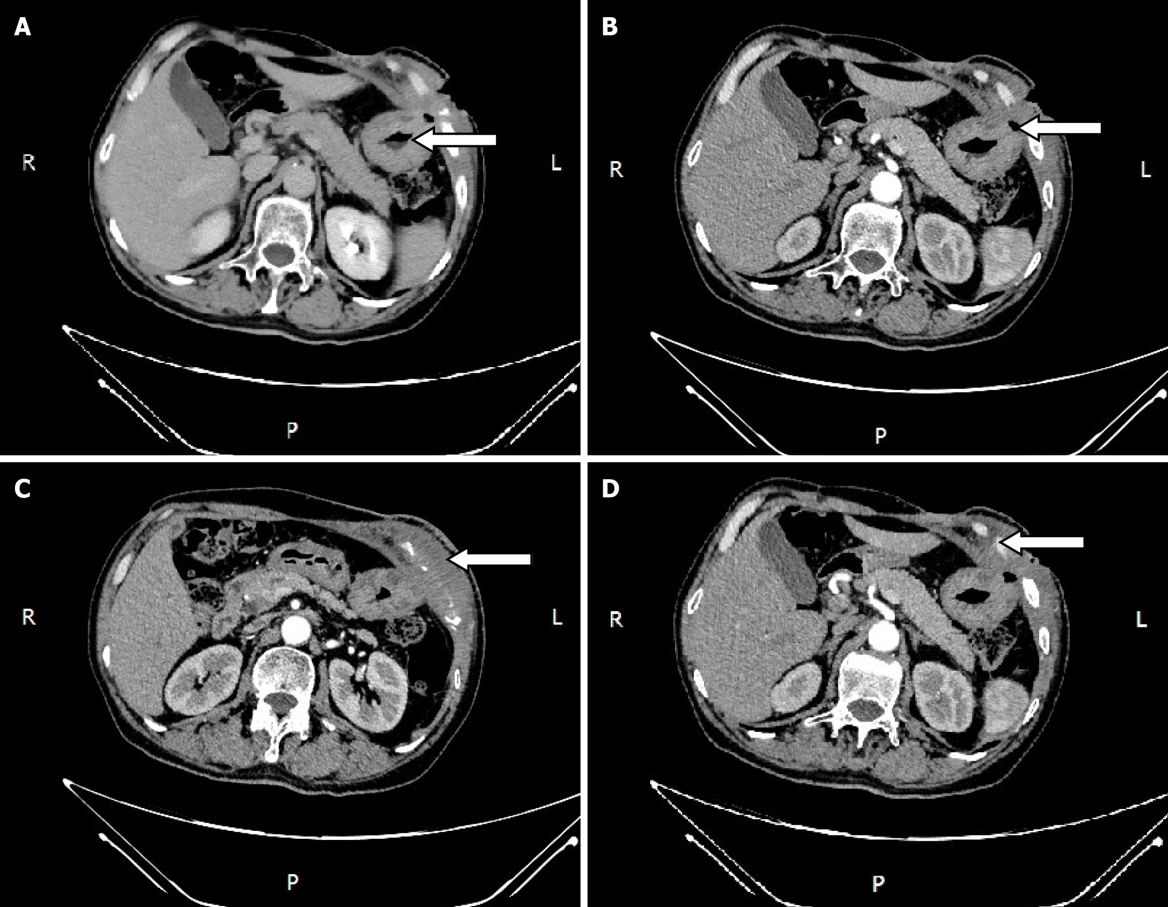
Figure 3 Abdominal enhanced computed tomography scan findings.
A: A thick gastric wall, and local discontinuity of the gastric wall in the greater curvature of the anterior gastric wall (arrow); B: A gas shadow in the gastric cavity also formed an outer bulge (arrow); C: Soft tissue swelling of thoracoabdominal wall (arrow); D: Rib destruction (arrow).
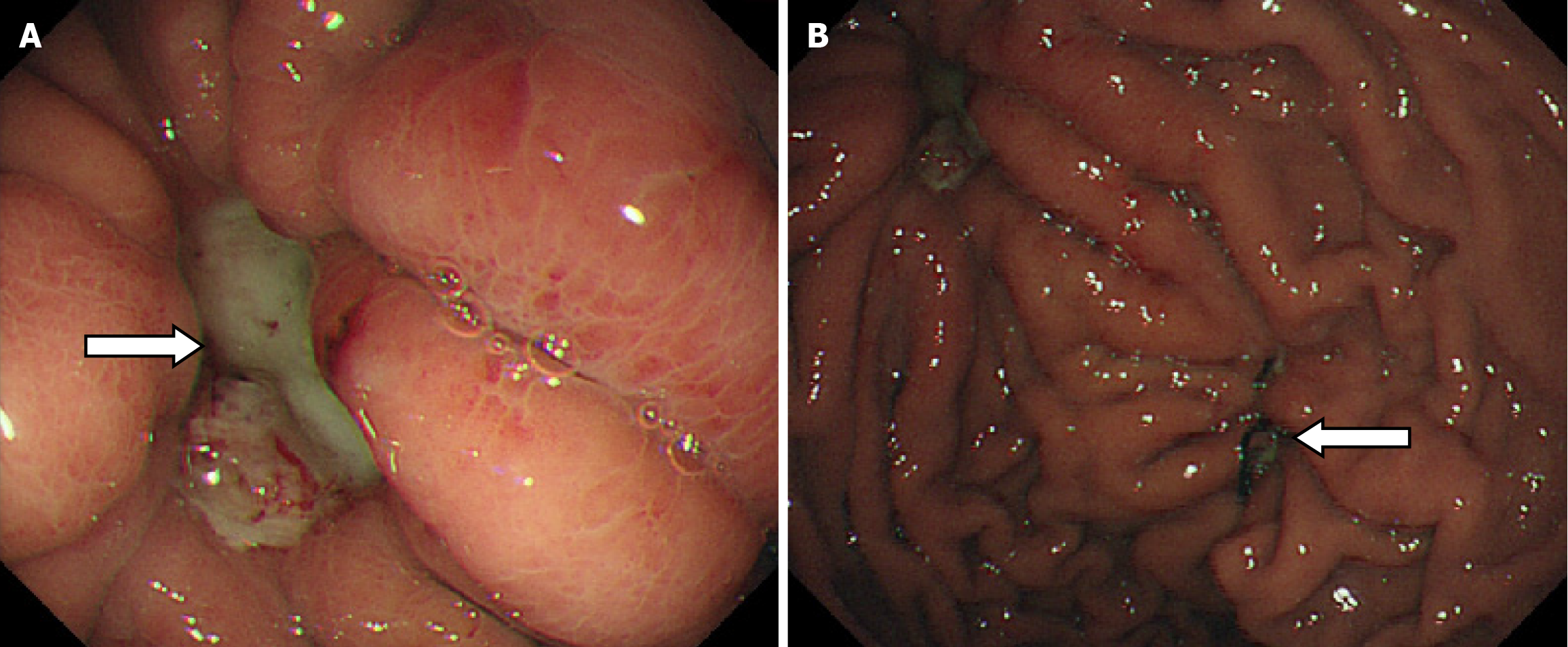
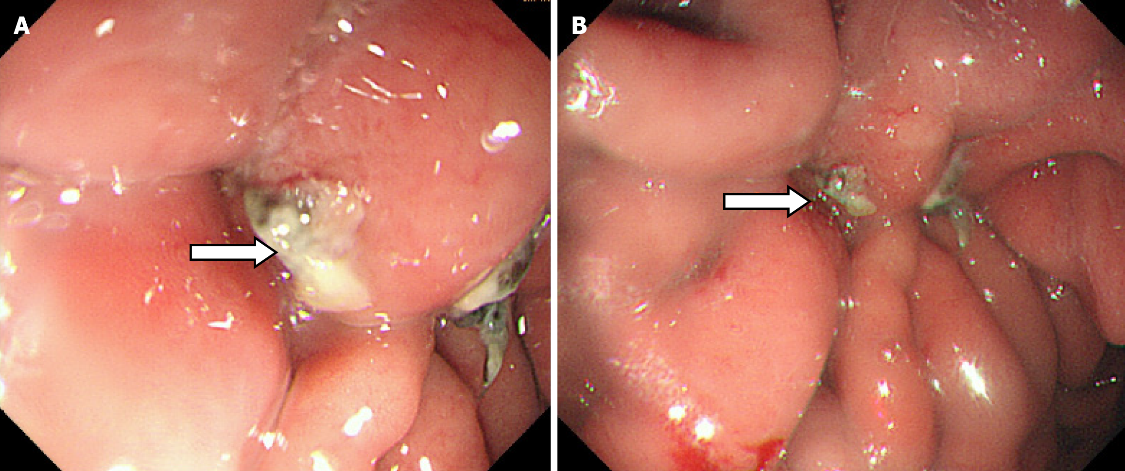
Figure 4 Gastric endoscopy showed a gastric ulcer.
A: A 1.8 cm × 1.2 cm deep ulcer on the anterior wall of the gastric body, with thick yellow material and hyperplastic tissue at the edge (arrow); B: The original surgical suture line was visible (arrow).
Figure 5 Abdominal computed tomography scan showed improvement in thoracoabdominal wall swelling.
Abdominal computed tomography scan showed (white arrow) improvement in thoracoabdominal wall swelling.
Figure 6 Gastric ulcer, stage A2.
A and B: Gastric body: Mucosal blotchy congestion and erosion, a 1.5 cm × 0.8 cm ulcer with white material and a clear mucus pool on the anterior wall of the greater curvature. Arrow: Ulcer.
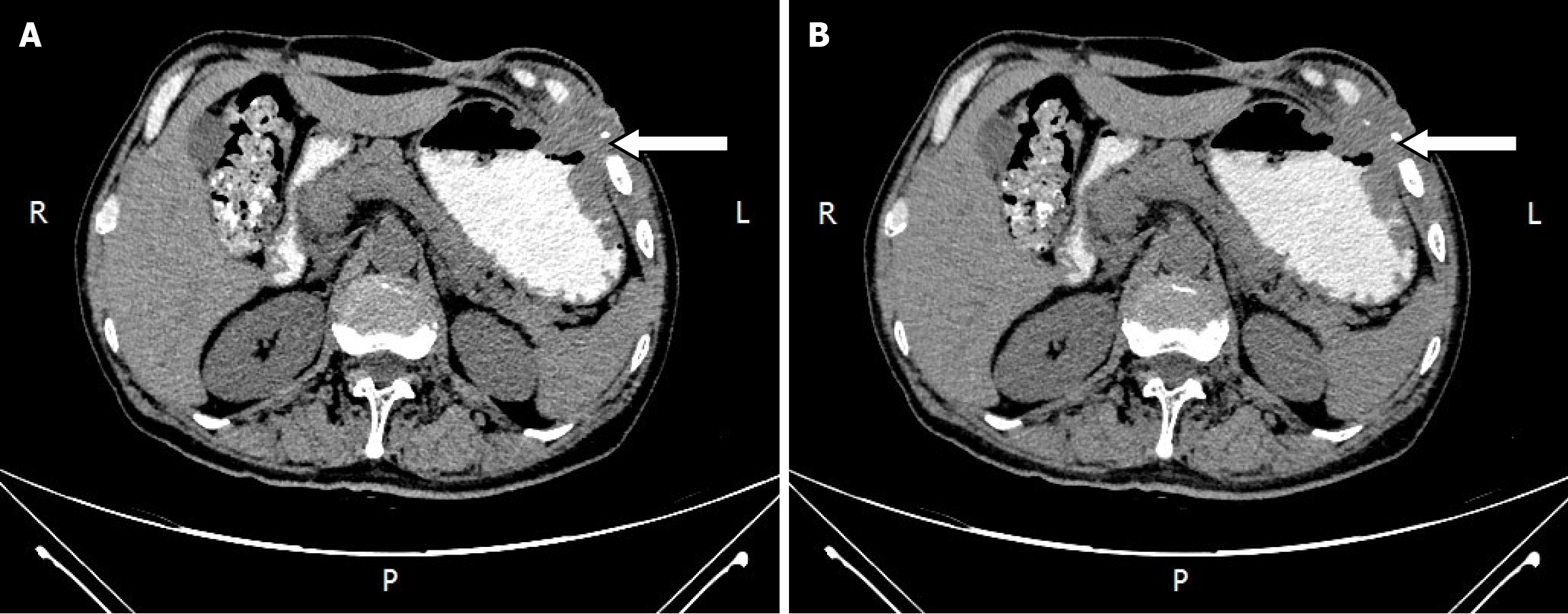
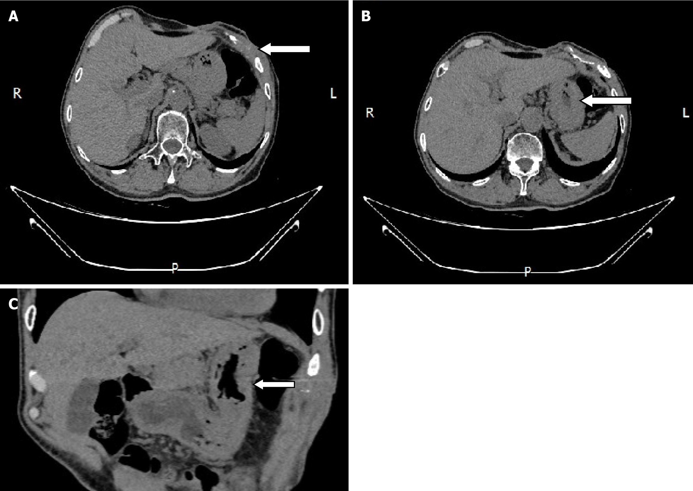
Figure 7 Repeated abdominal computed tomography on January 7, 2021 showed complete recovery.
A: Healed chest wall (arrow); B: Healed stomach wall (axial plane) (arrow); C: Healed stomach wall (coronal plane) (arrow).
Figure 8
Timeline about diagnosis and treatment of the patient.
- Citation: Kang YZ, Sun JH. Chronic gastro-abdominal wall fistula with secondary massive thoracoabdominal wall abscess and costal destruction after laparoscopic gastricolithotomy: A case report. World J Gastrointest Surg 2025; 17(6): 107033
- URL: https://www.wjgnet.com/1948-9366/full/v17/i6/107033.htm
- DOI: https://dx.doi.org/10.4240/wjgs.v17.i6.107033


