Copyright
©The Author(s) 2025.
World J Gastrointest Surg. Jun 27, 2025; 17(6): 104652
Published online Jun 27, 2025. doi: 10.4240/wjgs.v17.i6.104652
Published online Jun 27, 2025. doi: 10.4240/wjgs.v17.i6.104652
Figure 1 Schematic techniques of pancreaticojejunostomy after pancreaticoduodenectomy.
A: External ductal stent: Duct-to-mucosa anastomosis and outer layer joining pancreatic parenchyma and jejunum; B: Internal ductal stent: Duct-to-mucosa anastomosis and outer layer joining pancreatic parenchyma and jejunum; C: No ductal stent: Invagination of the transected pancreas by pancreaticojejunostomy (inner layer) and outer layer joining pancreatic parenchyma and jejunum.
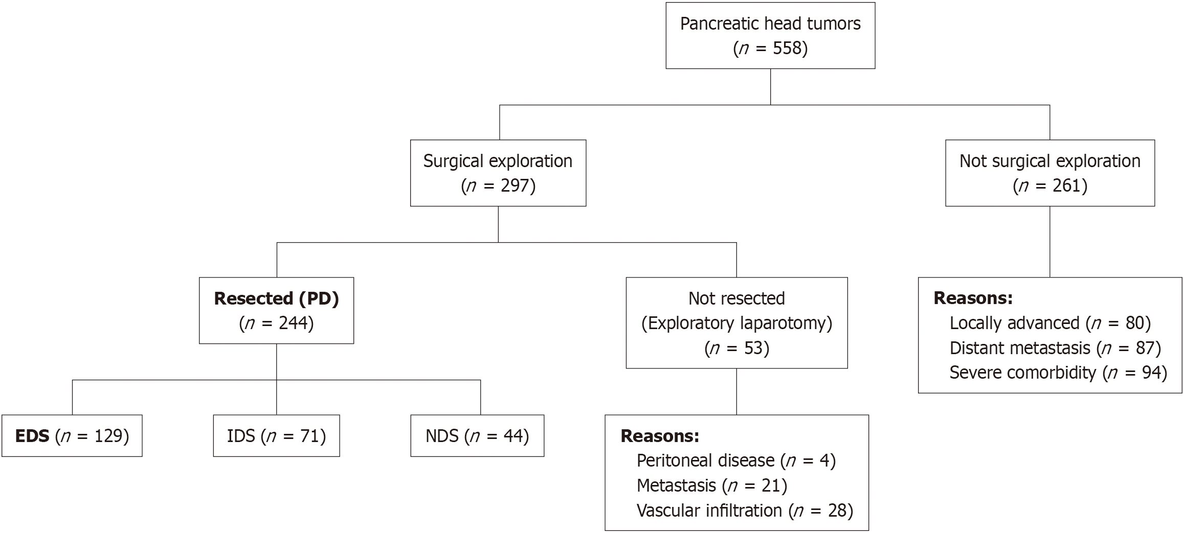
Figure 2 Flowchart of patient selection.
PD: Pancreatoduodenectomy; EDS: External duct stent; IDS: Internal duct stent; NDS: Non-ductal stent.
- Citation: Jiménez-Romero C, Marcacuzco-Quinto A, Caso-Maestro O, Alonso L, Fernández-Fernández C, Justo I. Comparison of three reconstruction techniques performed after pancreaticoduodenectomy: Using external, internal, or no stent. World J Gastrointest Surg 2025; 17(6): 104652
- URL: https://www.wjgnet.com/1948-9366/full/v17/i6/104652.htm
- DOI: https://dx.doi.org/10.4240/wjgs.v17.i6.104652


