Copyright
©The Author(s) 2021.
World J Gastrointest Surg. Jul 27, 2021; 13(7): 702-716
Published online Jul 27, 2021. doi: 10.4240/wjgs.v13.i7.702
Published online Jul 27, 2021. doi: 10.4240/wjgs.v13.i7.702
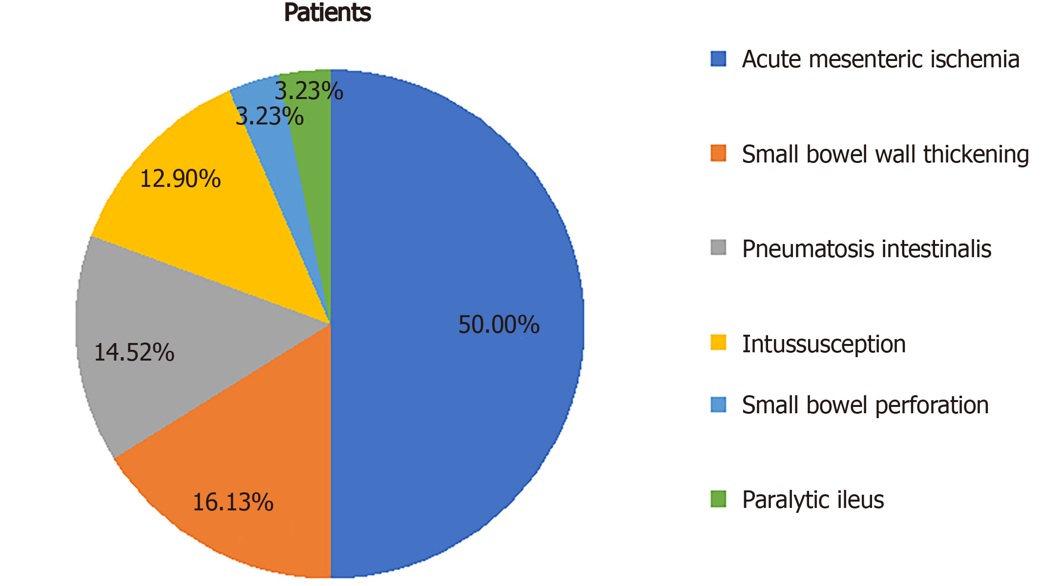
Figure 1 Proportions of radiologic small bowel manifestations reported since the beginning of the severe acute respiratory syndrome coronavirus 2 pandemic.
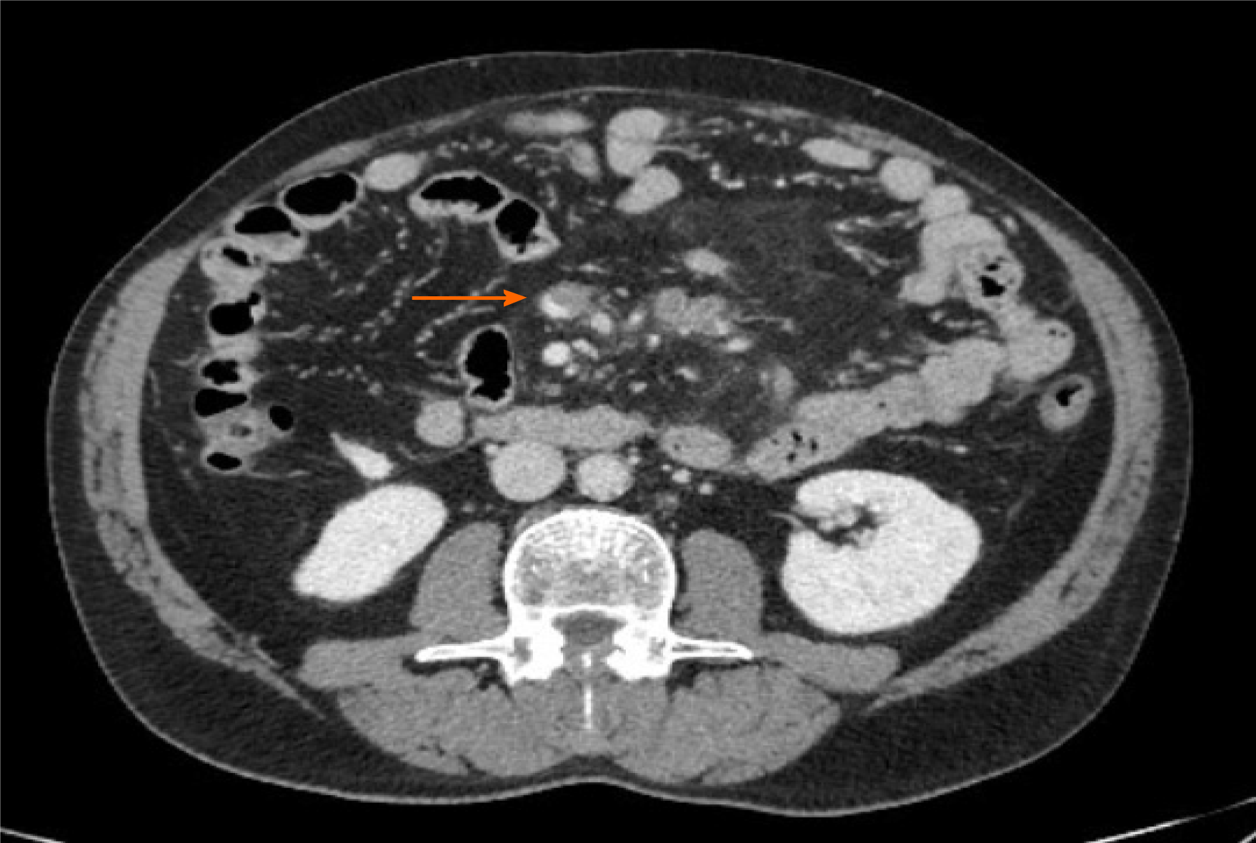
Figure 2 Computed tomography image showing partial superior mesenteric vein thrombosis in a man with severe acute respiratory syndrome coronavirus 2 infection.
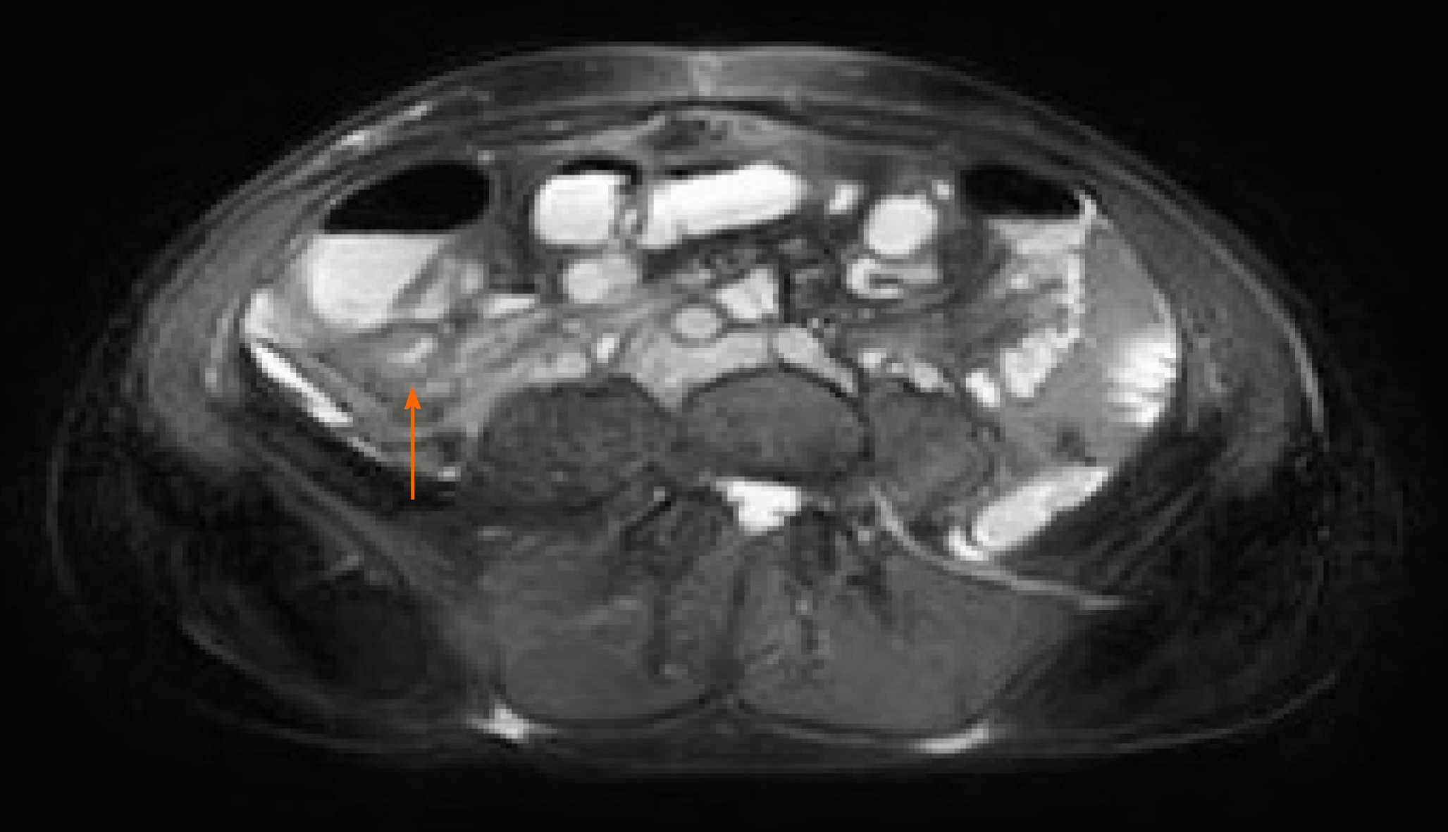
Figure 3 Magnetic resonance image showing distal ileum wall thickening in a young woman with severe acute respiratory syndrome coronavirus 2 infection.
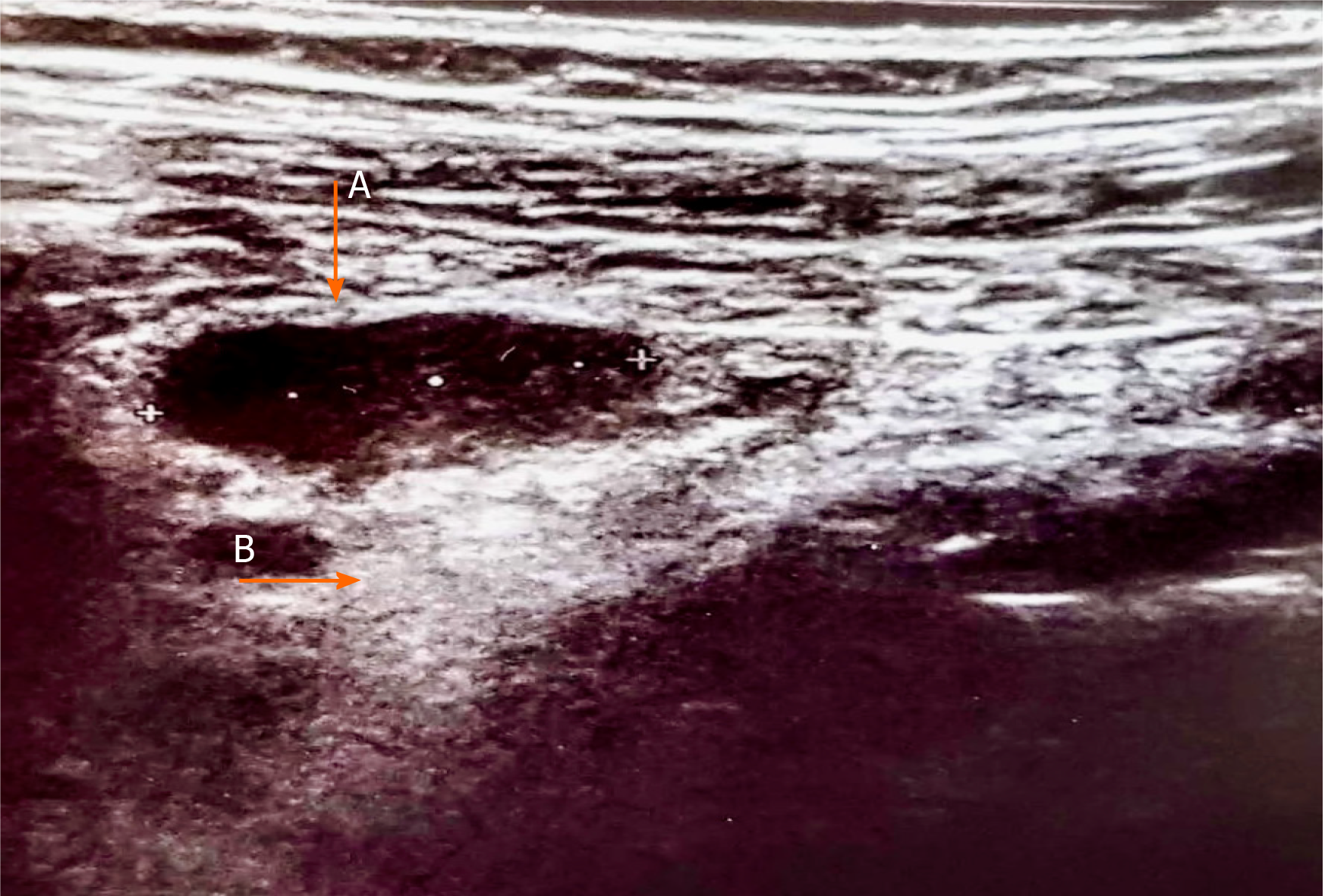
Figure 4 Abdominal ultrasound image in a 34-year-old woman with severe acute respiratory syndrome coronavirus 2 infection.
Enlarged hypoechoic mesenteric lymph nodes, with a maximum longitudinal axis diameter of 17 mm (arrow, A) and hyperechoic mesenteric adipose tissue hypertrophy (arrow, B) are shown.
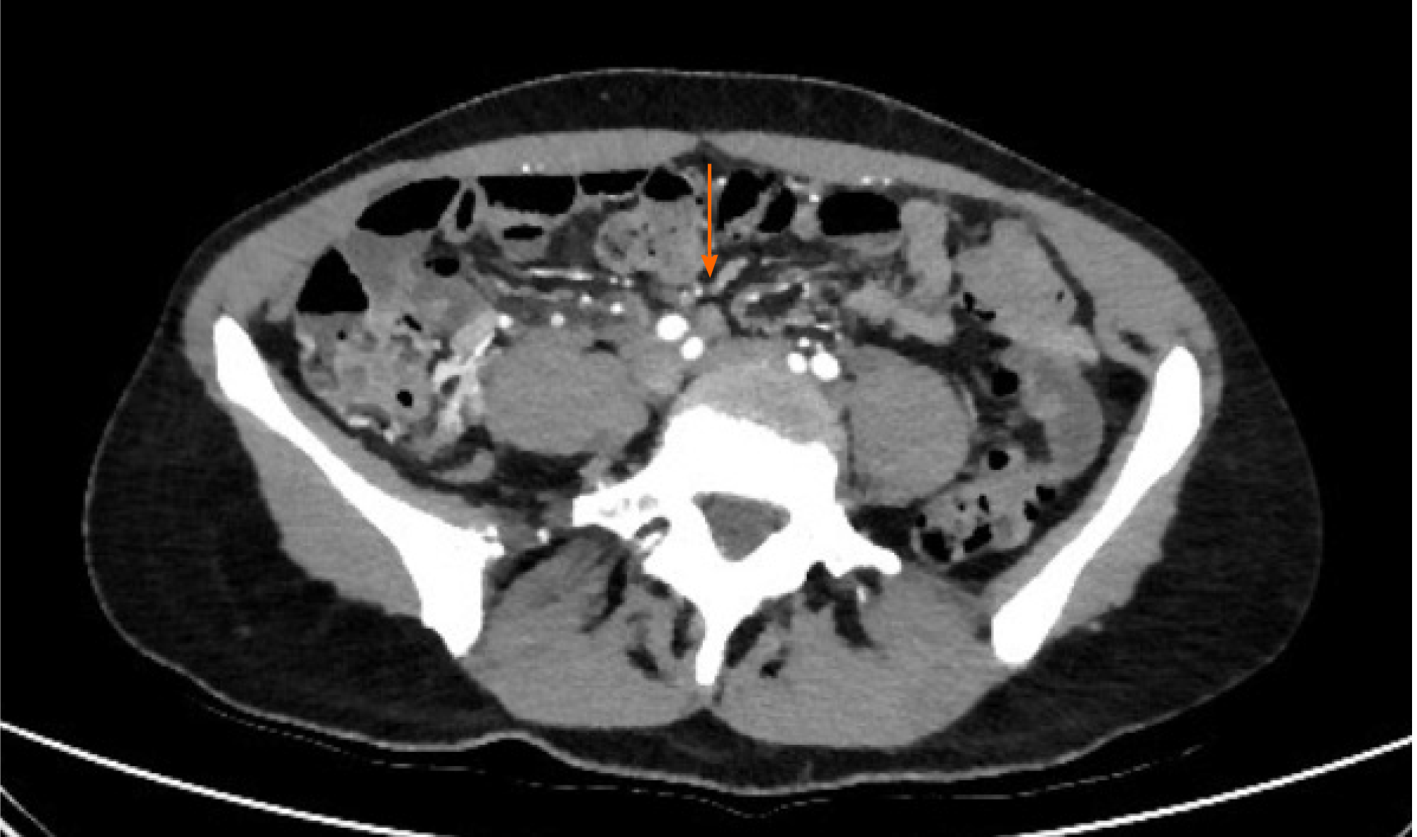
Figure 5 Abdominal computed tomography image showing multiple enlarged lymph nodes and mesenteric adipose tissue hypertrophy in a 34-year-old woman with severe acute respiratory syndrome coronavirus 2 infection.
- Citation: Pirola L, Palermo A, Mulinacci G, Ratti L, Fichera M, Invernizzi P, Viganò C, Massironi S. Acute mesenteric ischemia and small bowel imaging findings in COVID-19: A comprehensive review of the literature. World J Gastrointest Surg 2021; 13(7): 702-716
- URL: https://www.wjgnet.com/1948-9366/full/v13/i7/702.htm
- DOI: https://dx.doi.org/10.4240/wjgs.v13.i7.702


