Copyright
©The Author(s) 2021.
World J Gastrointest Surg. Feb 27, 2021; 13(2): 116-126
Published online Feb 27, 2021. doi: 10.4240/wjgs.v13.i2.116
Published online Feb 27, 2021. doi: 10.4240/wjgs.v13.i2.116
Figure 1 Procedure with a clutch cutter.
A: Opening and rotation of the clutch cutter; B: Horizontal cutting line; and C: Target tissue successfully grasped by the clutch cutter.
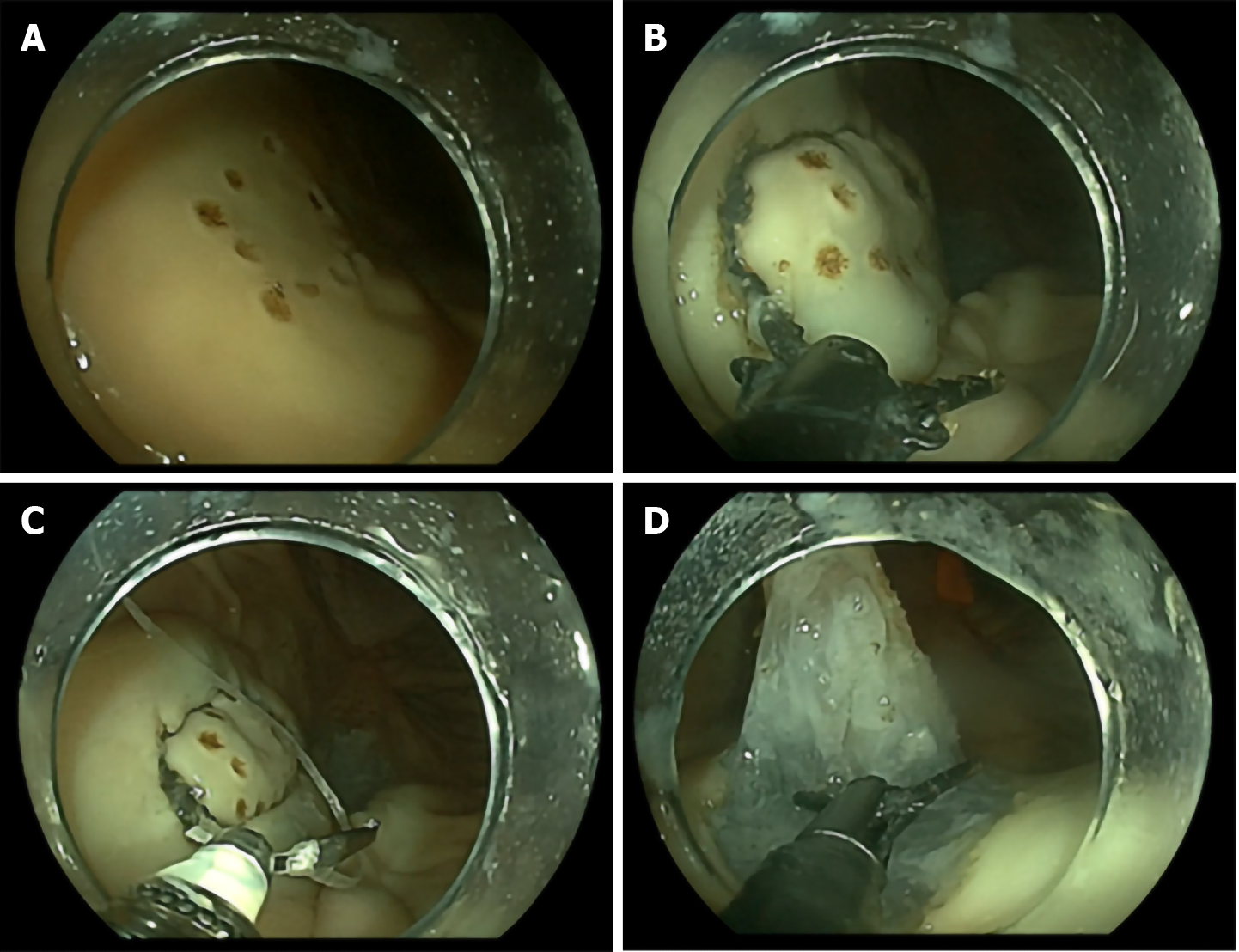
Figure 2 Endoscopic submucosal dissection procedure with a clutch cutter.
Representative images of each step of the endoscopic submucosal dissection with a clutch cutter procedure. A: Mock lesion made in the porcine stomach; B: Circumferential mucosal incision with a clutch cutter; C: Clip-with-thread traction method attached to the proximal edge of the lesion; and D: Submucosal dissection with a clutch cutter.
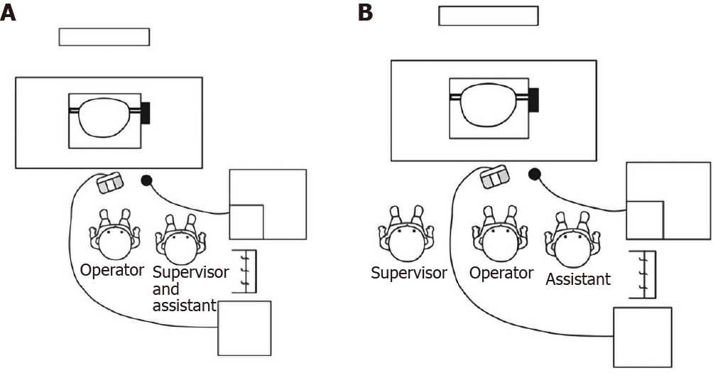
Figure 3 Positioning of the staff and instruments during endoscopic submucosal dissection with a clutch cutter.
A: Endoscopic submucosal dissection with a clutch cutter assisted by an expert; B: Endoscopic submucosal dissection with a clutch cutter assisted by a non-expert.
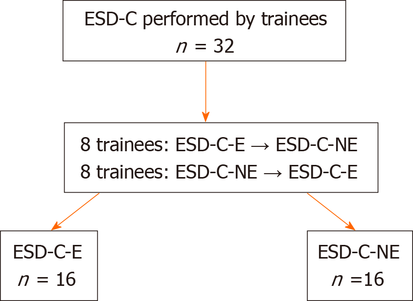
Figure 4 Flowchart of this study.
ESD-C: Endoscopic submucosal dissection with a clutch cutter; ESD-C-E: Endoscopic submucosal dissection with a clutch cutter assisted by an expert; ESD-C-NE: Endoscopic submucosal dissection with a clutch cutter assisted by a non-expert.
- Citation: Esaki M, Horii T, Ichijima R, Wada M, Sakisaka S, Abe S, Tomoeda N, Kitagawa Y, Nishioka K, Minoda Y, Tsuruta S, Suzuki S, Akiho H, Ihara E, Ogawa Y, Gotoda T. Assistant skill in gastric endoscopic submucosal dissection using a clutch cutter. World J Gastrointest Surg 2021; 13(2): 116-126
- URL: https://www.wjgnet.com/1948-9366/full/v13/i2/116.htm
- DOI: https://dx.doi.org/10.4240/wjgs.v13.i2.116


