Copyright
©The Author(s) 2020.
World J Gastrointest Surg. Mar 27, 2020; 12(3): 116-122
Published online Mar 27, 2020. doi: 10.4240/wjgs.v12.i3.116
Published online Mar 27, 2020. doi: 10.4240/wjgs.v12.i3.116
Figure 1 Thyroid gland hypermetabolism on positron emission tomography.
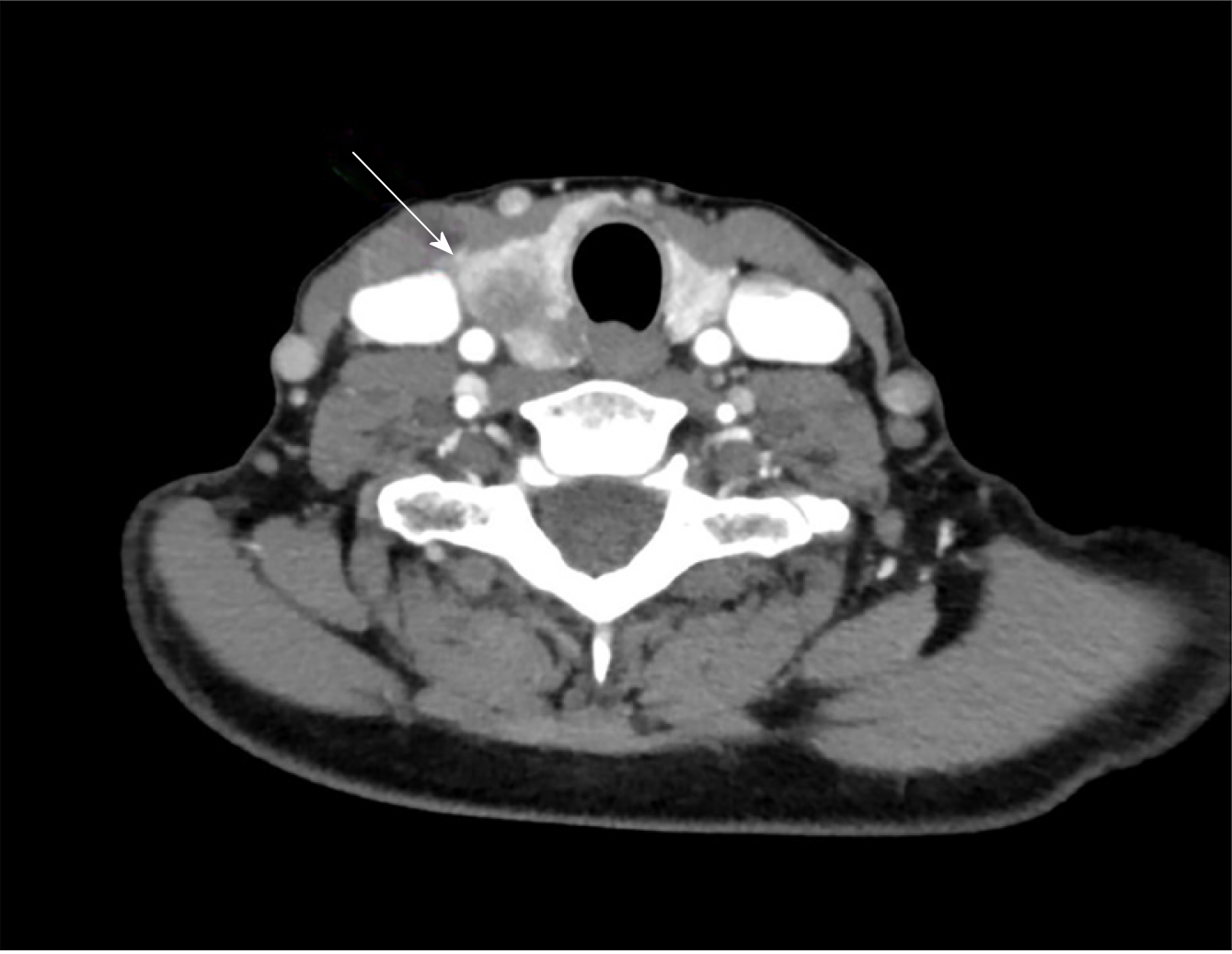
Figure 2 Computerized tomography image of a suspicious thyroid nodule.
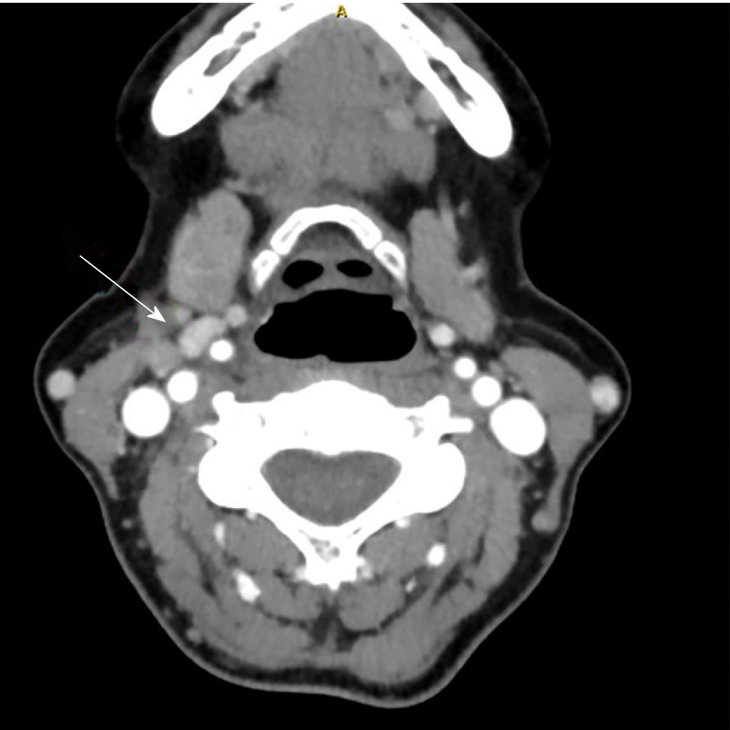
Figure 3 Computerized tomography image of a pericarotid lymph node.
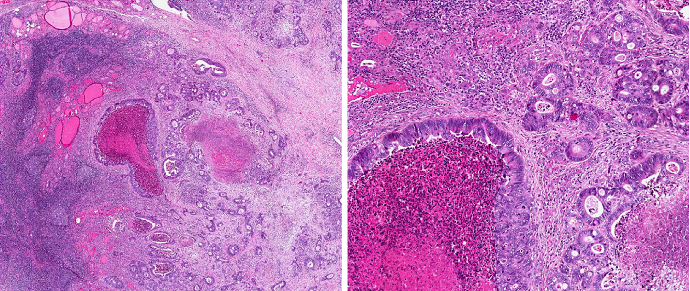
Figure 4 Hematoxylin-eosin stain of a specimen showing thyroid gland infiltration by metastasis of colorectal origin.
Numerous mitosis and necrosis (2.5 ×).
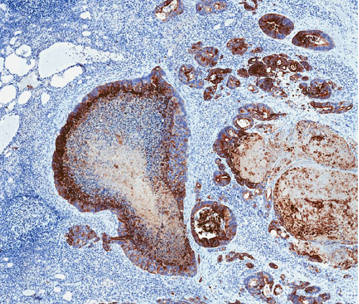
Figure 5 CK20-positive stain in a specimen (4.
5 ×).
- Citation: Ciriano Hernández P, Martínez Pinedo C, Calcerrada Alises E, García Santos E, Sánchez García S, Picón Rodríguez R, Jiménez Higuera E, Sánchez Peláez D, Herrera Montoro V, Martín Fernández J. Colorectal cancer metastases to the thyroid gland: A case report. World J Gastrointest Surg 2020; 12(3): 116-122
- URL: https://www.wjgnet.com/1948-9366/full/v12/i3/116.htm
- DOI: https://dx.doi.org/10.4240/wjgs.v12.i3.116


