Copyright
©The Author(s) 2019.
World J Gastrointest Surg. May 27, 2019; 11(5): 271-278
Published online May 27, 2019. doi: 10.4240/wjgs.v11.i5.271
Published online May 27, 2019. doi: 10.4240/wjgs.v11.i5.271
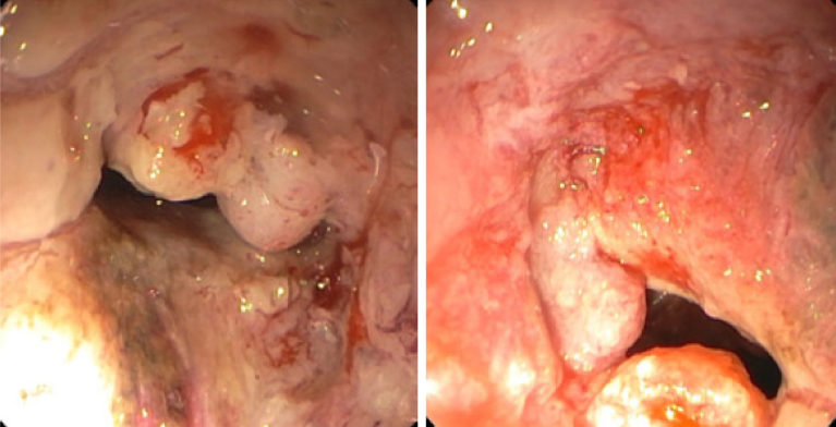
Figure 1 Rectal mass on colonoscopy.
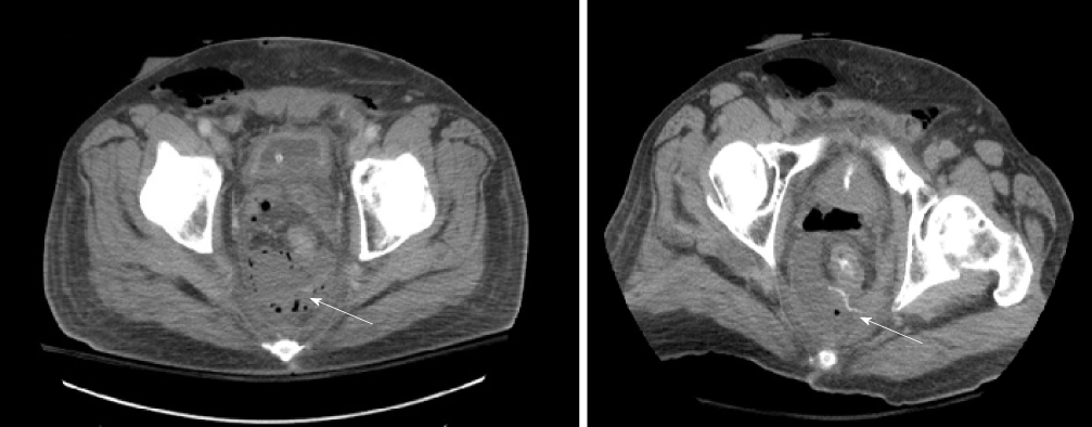
Figure 2 Presacral abscess with evidence of posterior leak of rectal contrast.
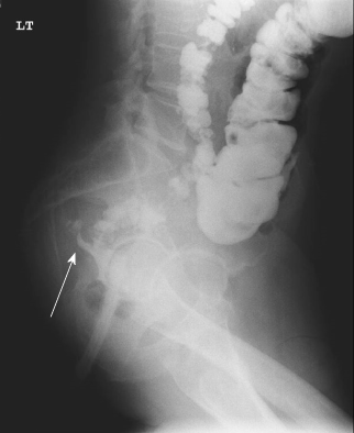
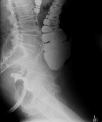
Figure 3 Posterior sinus tract evidence in barium contrast enema.
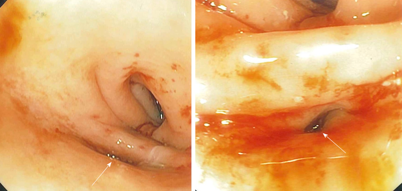
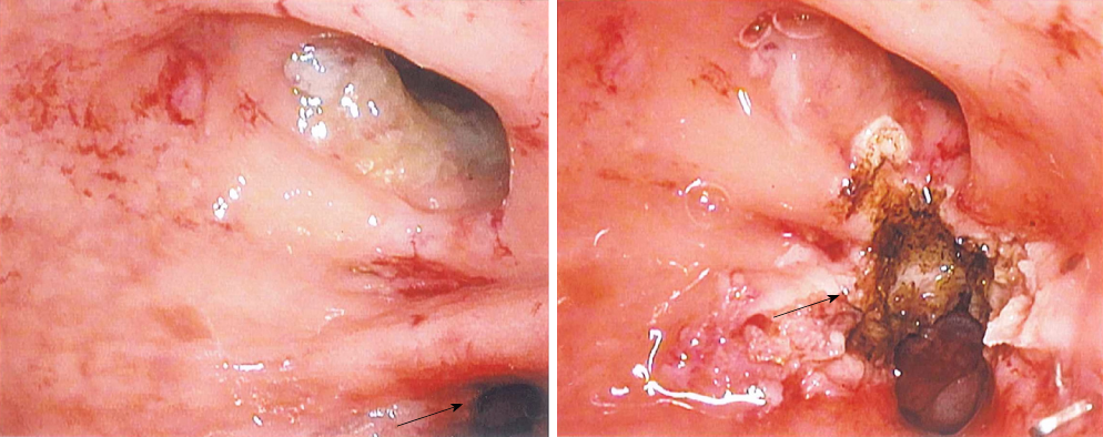
Figure 4 Posterior sinus tract at the healed staple line on flexible sigmoidoscopy.
Figure 5 Rectal lumen and posterior sinus tract and sinus tract after septotomy.
A: Rectal lumen and posterior sinus tract; B: Sinus tract after septotomy.
Figure 6 Absence of posterior sinus tract in post-operative barium contrast enema.
- Citation: Olavarria OA, Kress RL, Shah SK, Agarwal AK. Novel technique for anastomotic salvage using transanal minimally invasive surgery: A case report. World J Gastrointest Surg 2019; 11(5): 271-278
- URL: https://www.wjgnet.com/1948-9366/full/v11/i5/271.htm
- DOI: https://dx.doi.org/10.4240/wjgs.v11.i5.271


