Copyright
©The Author(s) 2015.
World J Diabetes. May 15, 2015; 6(4): 576-582
Published online May 15, 2015. doi: 10.4239/wjd.v6.i4.576
Published online May 15, 2015. doi: 10.4239/wjd.v6.i4.576
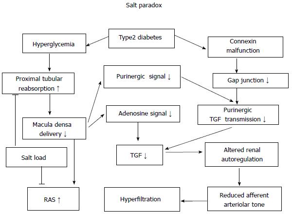
Figure 1 Working hypothesis for glomerular hyperfiltration in diabetes.
On the one hand, hyperglycemia enhances sodium reabsorption in type1 and type 2 diabetes, thereby decreasing the delivery to macula densa with resultant weakening of tubuloglomerular feedback (TGF). The latter impairs renal autoregulation that dilats afferent arterioles, and activates renin-angiotensin system (RAS). On the other hand, TGF signal by adenosine triphosphate (P2) is damaged in type 2 diabetes due to connexin phosphorylation and gap junction malfunction, worsening glomerular hyperifiltration. High salt intake inhibits proximal tubular reabsorption, thereby increasing the delivery of sodium chloride to macula densa. This ameliorates pathological afferent arteriolar dilation by the restoration of TGF through adenosine (A1) signal[18].
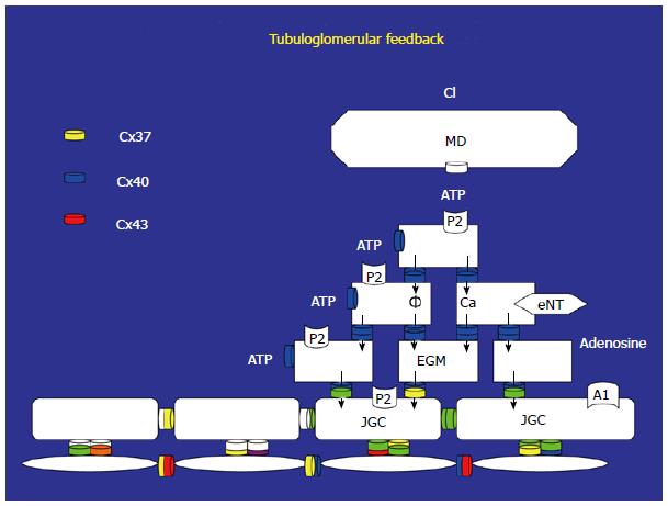
Figure 2 Sodium chloride is delivered to macula densa, macula densa releases adenosine triphosphate.
Adenosine triphosphate (ATP) binds to P2 receptor on extraglomerular mesangial cell (EGM), and induces membrane depolarization and/or elevations of cytosolic calcium. These signals are transduced to juxtaglomerular cells (JGC) by intercellular communication through gap junctions consisted of connexins (Cx). In addition, ATP is degraded to adenosine by ecto-nucleotidase (eNT) on EGM. Adenosine binds to A1 receptor on JGC, increasing cytosolic calcium. Calcium waves generated in JGC transduce through gap junctions between afferent arteriolar myocytes to its upstream, eliciting ascending vasoconsitiction (refs 8 and 10). MD: Macula densa; Cl: Chloride.
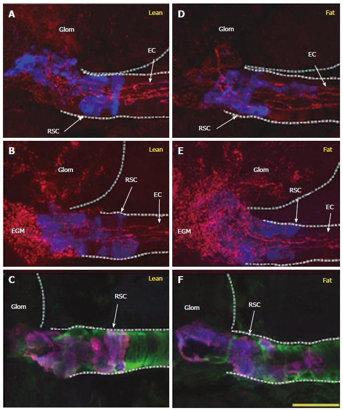
Figure 3 Expression of cennexins (Cx37, 40, 43) in juxtaglomerular apparatus of type 2 diabetic (Zucker diabetic fatty) and control (Zucker lean) rats.
A, B, D, E: Renin secreting cells (RSC, blue) and endothelial cells (EC) express Cx37 (A: Lean; D: Fat) and Cx40 (B: Lean; E: Fat). Cx40 is expressed on glomerular and extraglomerular mesangial cells (EGM) in control (B) and diabetes (E). Cx43 is expressed in cytosol of RSC in both groups (C, F). Quantification reveals that the expression of Cx37 in RSC is reduced in diabetic model[15].
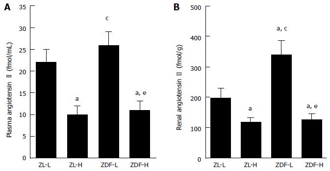
Figure 4 Plasma and kidney concentration of angiotensin II in type 2 diabetic model (Zucker diabetic fatty rat) and control rat (Zucker lean rat).
ZL-L and ZL-H indicate ZL fed normal and high salt diet, respectively. Alike, ZDF-L, ZDF-H describe ZDF fed normal and high salt diet, respectively. aP < 0.05 vs ZL-L, cP < 0.05 vs ZL-H, eP < 0.05 vs ZDF-L[18]. ZDF: Zucker diabetic fatty rat; ZL: Zucker lean rat.
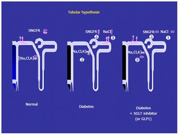
Figure 5 Compared to normal condition (left), ultrafiltrate in Bowman capsule contains significant amount of glucose in diabetes (middle).
Because proximal tubules reabsorb more sodium with glucose through SGLT in diabetes (1), the delivery to macula densa is decreased (2). This weakens TGF signals to increase glomerular filtration rate (3), accounting for hyperfiltration in early stage of diabetes. Either GLP or SGLT inhibition (right) inhibits proximal tubular reabsorption (1), restoring sodium chloride delivery to macula densa even under hyperglycemic condition (2). This would have TGF work and normalize glomerular filtration rate (3), ameliorating glomerular hyperfiltration. SGLT: Sodium glucose co-transporter; GLP: Glucagon-like peptide; TGF: Tubuloglomerular feedback.
- Citation: Takenaka T, Inoue T, Watanabe Y. How the kidney hyperfiltrates in diabetes: From molecules to hemodynamics. World J Diabetes 2015; 6(4): 576-582
- URL: https://www.wjgnet.com/1948-9358/full/v6/i4/576.htm
- DOI: https://dx.doi.org/10.4239/wjd.v6.i4.576


