Copyright
©The Author(s) 2015.
World J Diabetes. May 15, 2015; 6(4): 548-553
Published online May 15, 2015. doi: 10.4239/wjd.v6.i4.548
Published online May 15, 2015. doi: 10.4239/wjd.v6.i4.548
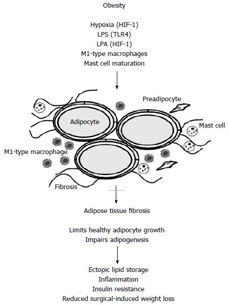
Figure 1 In obese adipose tissue hypoxia, lipopolysaccharide, lysophosphatidic acid, M1-type macrophages and maturated mast cells contribute to adipose tissue fibrogenesis.
Hypoxia and LPA mediated effects involve activation of HIF-1 and LPS activates TLR4. Fibrosis impairs adipogenesis and healthy adipocyte growth. Lipids are therefore stored in peripheral tissues like the liver. This is associated with impaired insulin sensitivity and inflammation. Adipose tissue fibrosis negatively affects surgery-induced weight loss. LPS: Lipopolysaccharide; LPA: Lysophosphatidic acid; HIF-1: Hypoxia inducible factor 1.
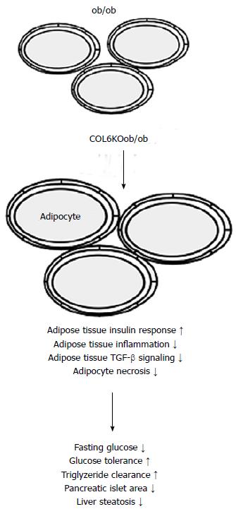
Figure 2 Mice with leptin deficiency (ob/ob mice) have large adipocytes and their size further increases when collagen 6 (COL6KOob/ob) is knocked-out in these animals.
Weakening of the extracellular matrix is associated with improved adipose tissue insulin response, reduced inflammation and TGF-beta signaling, and diminished adipocyte necrosis. Subsequently metabolic situation is improved.
- Citation: Buechler C, Krautbauer S, Eisinger K. Adipose tissue fibrosis. World J Diabetes 2015; 6(4): 548-553
- URL: https://www.wjgnet.com/1948-9358/full/v6/i4/548.htm
- DOI: https://dx.doi.org/10.4239/wjd.v6.i4.548


