Copyright
©2014 Baishideng Publishing Group Inc.
World J Diabetes. Dec 15, 2014; 5(6): 756-762
Published online Dec 15, 2014. doi: 10.4239/wjd.v5.i6.756
Published online Dec 15, 2014. doi: 10.4239/wjd.v5.i6.756
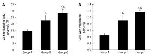
Figure 1 Percentage of apoptotic and dead cells in healthy (Group A), type 2 diabetes mellitus (Group B) and type 2 diabetes mellitus patients with chronic non healing wound (Group C) (A and B).
bP < 0.01 vs healthy; aP < 0.05 vs uncontrolled diabetes without complication and uncontrolled diabetes with chronic non healing wound. First, second, and third bar in each panel represents healthy, uncontrolled diabetic and uncontrolled diabetic with chronic non healing wound, respectively.
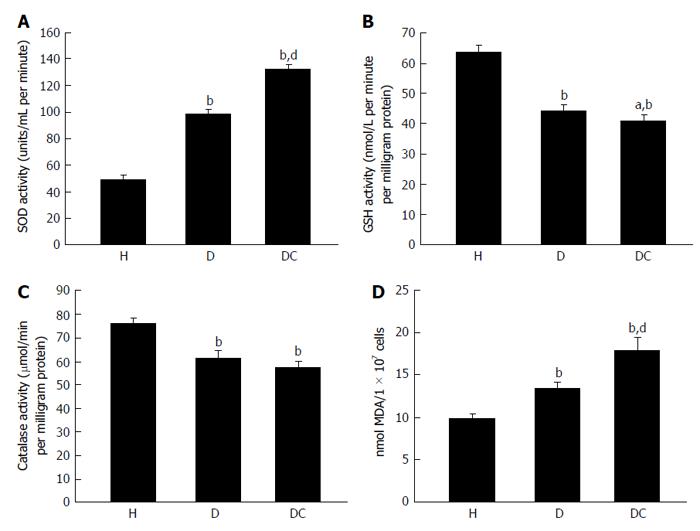
Figure 2 Concentration of superoxide dismutase (A), reduced glutathione (B), catalase (C) and malondialdehyde (D) in healthy (H), type 2 diabetes mellitus (D) and type 2 diabetes mellitus patients with chronic non healing (DC) groups.
bP < 0.01 vs healthy; dP < 0.01 and aP < 0.05 vs uncontrolled diabetes without complication and uncontrolled diabetes with chronic non healing wound. First, second, and third bar in each panel represents healthy, uncontrolled diabetic and uncontrolled diabetic with chronic non healing wound, respectively. SOD: Superoxide dismutase; GSH: Reduced glutathione; MDA: Malondialdehyde.
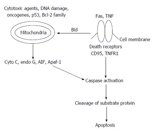
Figure 3 Basic outline of apoptosis mechanism.
Bcl-2: B-cell lymphoma 2; TNF: Tumor necrosis factor; AIF: Apoptosis-inducing factor; Apaf-1: Apoptotic protease activating factor-1; TNFR1: Tumor necrosis factor receptor 1.
- Citation: Arya AK, Tripathi R, Kumar S, Tripathi K. Recent advances on the association of apoptosis in chronic non healing diabetic wound. World J Diabetes 2014; 5(6): 756-762
- URL: https://www.wjgnet.com/1948-9358/full/v5/i6/756.htm
- DOI: https://dx.doi.org/10.4239/wjd.v5.i6.756


