Copyright
©2014 Baishideng Publishing Group Inc.
World J Diabetes. Aug 15, 2014; 5(4): 482-492
Published online Aug 15, 2014. doi: 10.4239/wjd.v5.i4.482
Published online Aug 15, 2014. doi: 10.4239/wjd.v5.i4.482
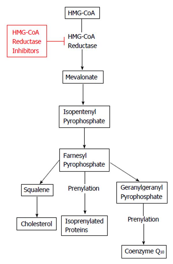
Figure 1 Effect of HMG-CoA reductase inhibitors (statins) on cholesterol and coenzyme Q10.
HMG-CoA: 3-hydroxy-3-methylglutaryl-coenzyme A.
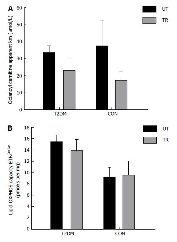
Figure 2 Patients with type 2 diabetes (n = 5) and healthy control subjects (n = 5) performed eight sessions of one-legged high intensity training in two weeks.
Each session consisted of ten one-minute exercise bouts at 60% of one-legged maximal oxygen uptake and > 80% of maximal heart rate, interspersed by one min rest. After completion of the training muscle biopsies (vastus lateralis) were obtained from the untrained (black bars) and the trained (grey bars) leg. The measurement mitochondrial OXPHOS capacity and substrate sensitivity was performed with malate, ADP and octanoyl carnitine (titration: 5-2000 μmol/L). A: Apparent Michaelis Menten constant Km for octanoyl carnitine; B: Maximal OXPHOS capacity with the mentioned substrates. T2DM: Type 2 diabetes; CON: Control subjects; UT: Untrained; TR: Trained.
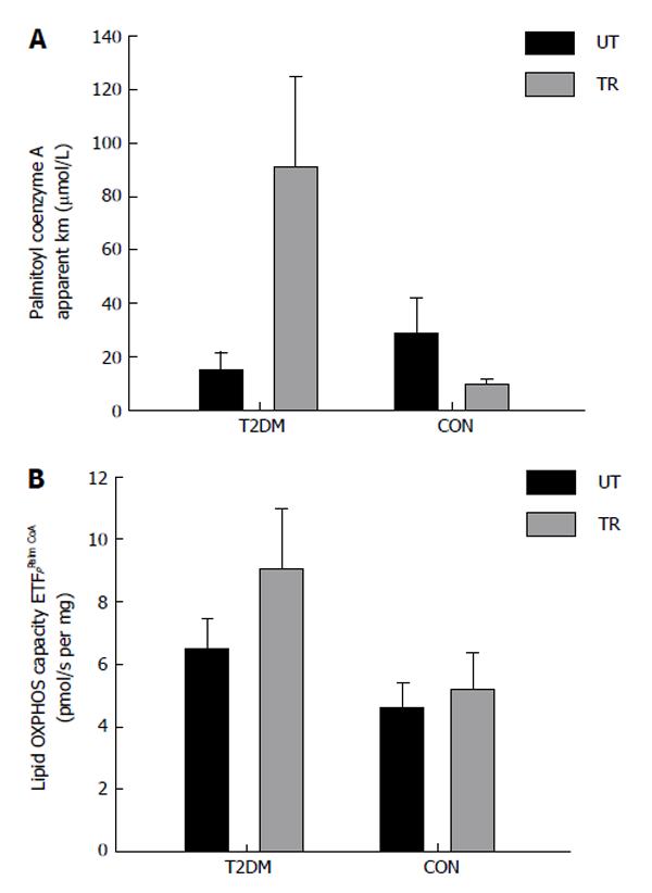
Figure 3 Patients with type 2 diabetes (n = 5) and healthy control subjects (n = 3) performed eight sessions of one-legged high intensity training in two weeks.
Each session consisted of ten one-minute exercise bouts at 60% of one-legged maximal oxygen uptake and > 80% of maximal heart rate, interspersed by one min rest. After completion of the training muscle biopsies (vastus lateralis) were obtained from the untrained (black bars) and the trained (grey bars) leg. The measurement mitochondrial OXPHOS capacity and substrate sensitivity was performed with malate, ADP and palmitoyl coenzyme A (titration: 5-100 μmol/L). A: Apparent Michaelis Menten constant Km for palmitoyl coenzyme A; B: Maximal OXPHOS capacity with the mentioned substrates. T2DM: Type 2 diabetes; CON: Control subjects; UT: Untrained; TR: Trained.
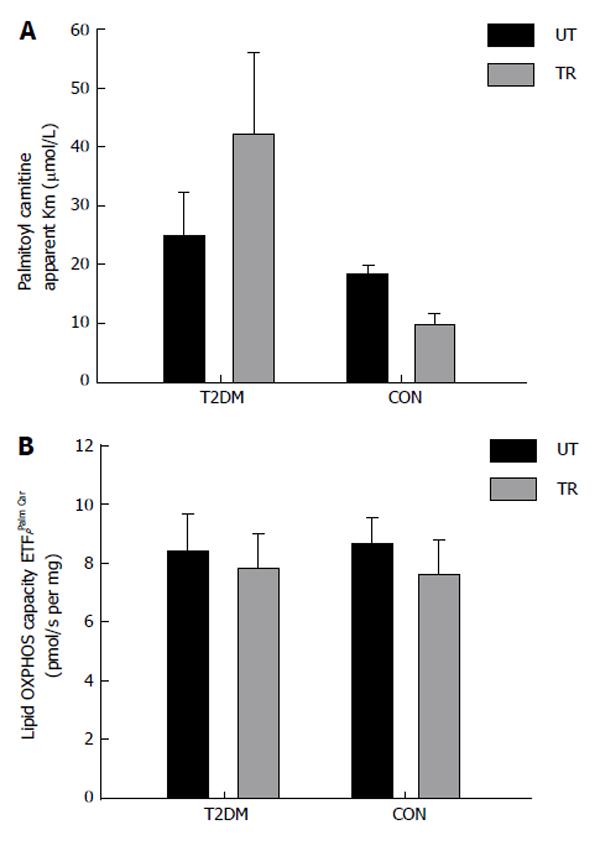
Figure 4 Patients with type 2 diabetes (n = 7) and healthy control subjects (n = 5) performed eight sessions of one-legged high intensity training in two weeks.
Each session consisted of ten one-minute exercise bouts at 60% of one-legged maximal oxygen uptake and > 80% of maximal heart rate, interspersed by one min rest. After completion of the training muscle biopsies (vastus lateralis) were obtained from the untrained (black bars) and the trained (grey bars) leg. The measurement of mitochondrial OXPHOS capacity and substrate sensitivity was performed with malate, ADP and palmitoyl carnitine (titration: 5-200 μmol/L). A: Apparent Michaelis Menten constant Km for palmitoyl carnitine; B: Maximal OXPHOS capacity with the mentioned substrates. T2DM: Type 2 diabetes; CON: Control subjects; UT: Untrained; TR: Trained.
- Citation: Larsen S, Skaaby S, Helge JW, Dela F. Effects of exercise training on mitochondrial function in patients with type 2 diabetes. World J Diabetes 2014; 5(4): 482-492
- URL: https://www.wjgnet.com/1948-9358/full/v5/i4/482.htm
- DOI: https://dx.doi.org/10.4239/wjd.v5.i4.482


