Copyright
©The Author(s) 2021.
World J Diabetes. Feb 15, 2021; 12(2): 158-169
Published online Feb 15, 2021. doi: 10.4239/wjd.v12.i2.158
Published online Feb 15, 2021. doi: 10.4239/wjd.v12.i2.158
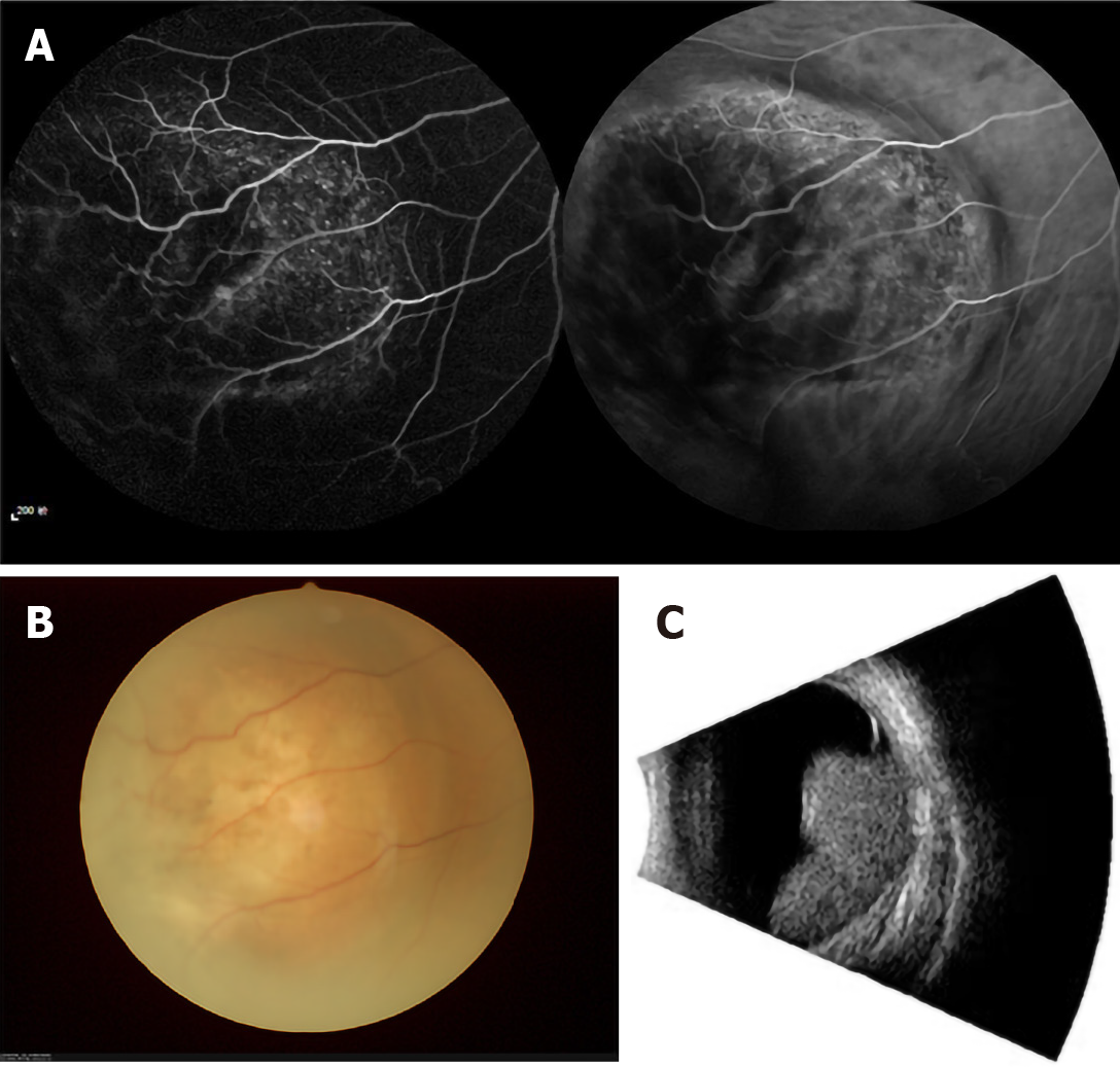
Figure 1 Example of patients with intraocular metastasis seen on fluorescence fundus angiography, fundus camera and eye B ultrasound, respectively.
A: Fluorescence fundus angiography; B: Fundus camera; C: Eye B ultrasound.
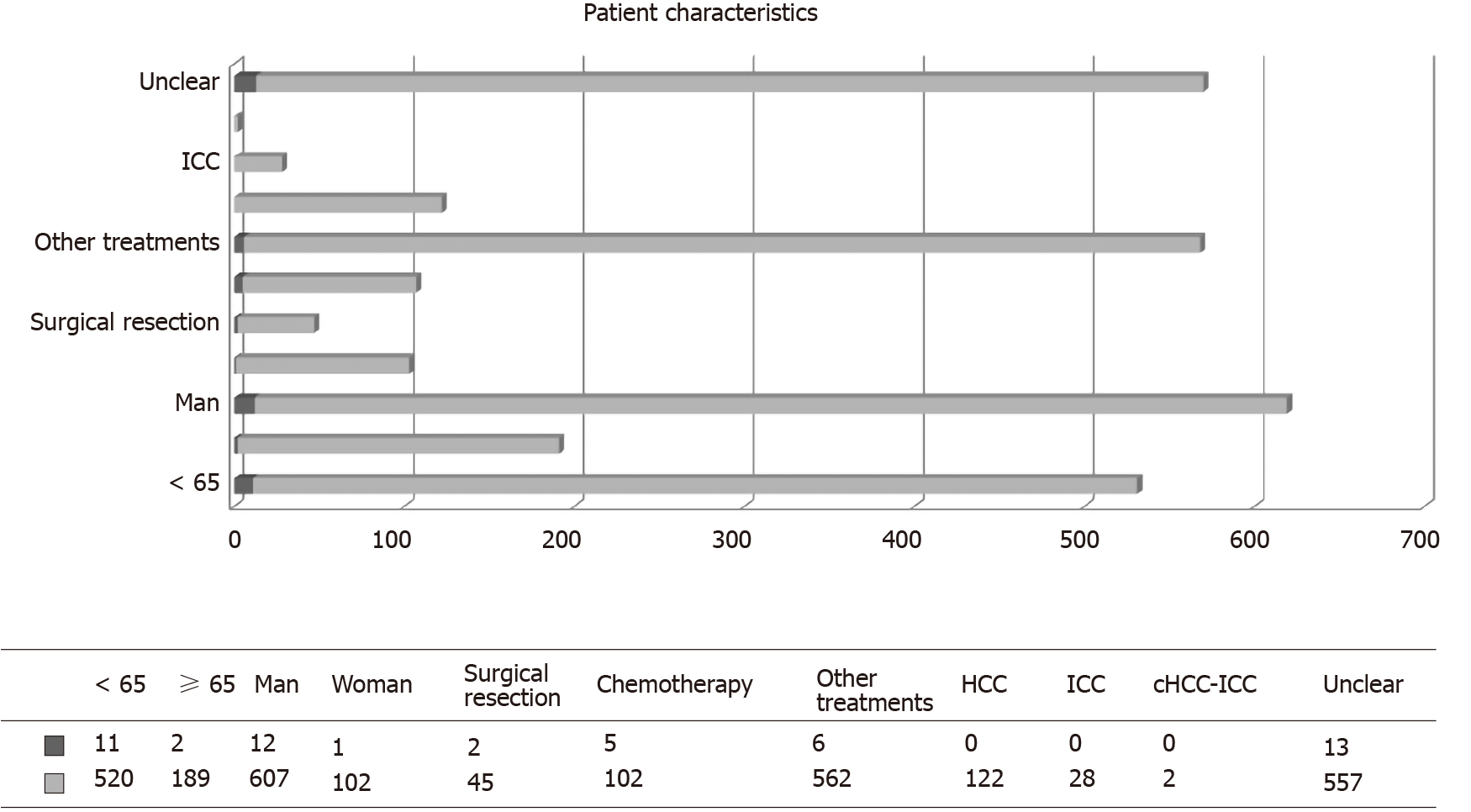
Figure 2 Clinical features of patients with and without intraocular metastasis.
Blue represents age, sex, treatments, and histopathological type in the intraocular metastasis group; red represents age, sex, treatment, and histopathological type in the non-intraocular metastasis group. “Unclear” means that the histopathological type is unclear. HCC: Hepatocellular carcinoma; ICC: Intrahepatic cholangiocarcinoma; cHCC-ICC: Combined hepatocellular carcinoma and intrahepatic cholangiocarcinoma.
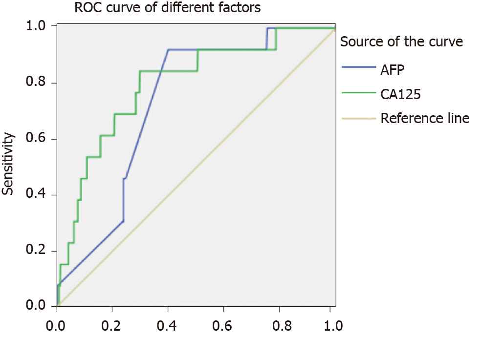
Figure 3 The receiver operating characteristic curves of risk factors for detecting intraocular metastasis in primary liver cancer.
ROC: Receiver operating characteristic; AFP: Alpha-fetoprotein; CA125: Cancer antigen 125.
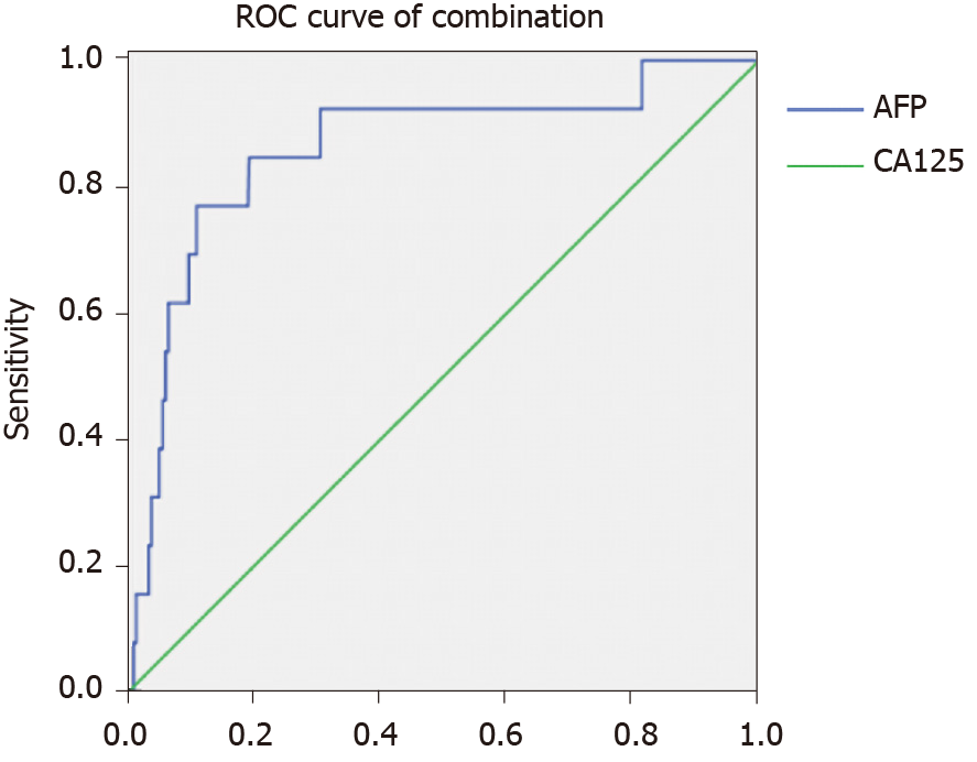
Figure 4 The receiver operating characteristic curves of the combination of alpha-fetoprotein and cancer antigen 125.
ROC: Receiver operating characteristic; AFP: Alpha-fetoprotein; CA125: Cancer antigen 125.
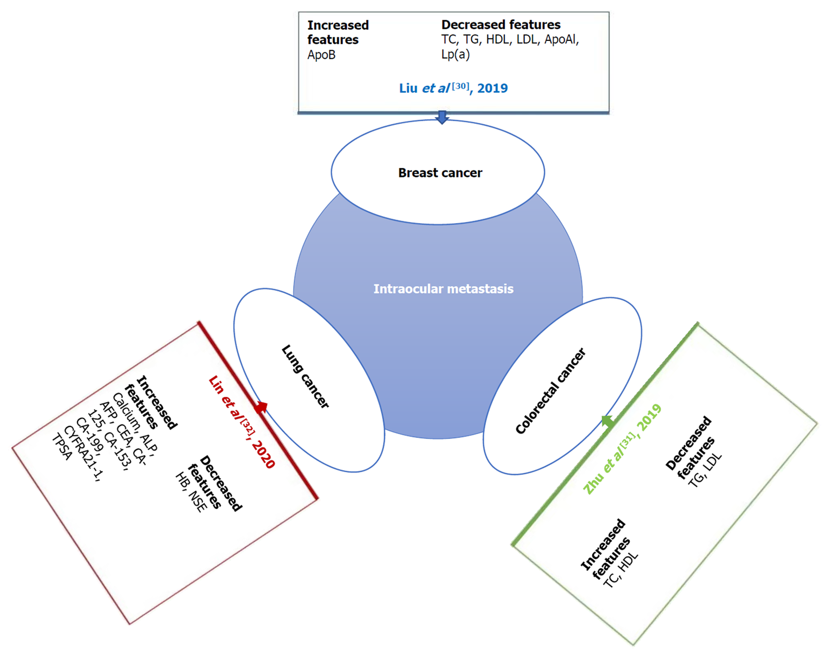
Figure 5 Studies on intraocular metastasis from different cancers.
Serological tumor markers were changed when different tumors had intraocular metastasis (IOM). The changes in serologic tumor markers during IOM of breast, lung, and colorectal cancer are shown here. ApoB: Apolipoprotein B; TC: Total cholesterol; TG: Triglyceride; HDL: High density lipoprotein; LDL: Low density lipoprotein; ApoA1: Apolipoprotein A1; Lp(a): Lipoprotein A; ALP: Alkaline phosphatase; AFP: Alpha-fetoprotein; CEA: Carcinoembryonic antigen; CA: Cancer antigen; CYFRA: Cytokeratin fragment; TPSA: Total prostate-specific antigen.
- Citation: Yu K, Tang J, Wu JL, Li B, Wu SN, Zhang MY, Li QY, Zhang LJ, Pan YC, Ge QM, Shu HY, Shao Y. Risk factors for intraocular metastasis of primary liver cancer in diabetic patients: Alpha-fetoprotein and cancer antigen 125. World J Diabetes 2021; 12(2): 158-169
- URL: https://www.wjgnet.com/1948-9358/full/v12/i2/158.htm
- DOI: https://dx.doi.org/10.4239/wjd.v12.i2.158


