Copyright
©2013 Baishideng Publishing Group Co.
World J Gastrointest Oncol. Jul 15, 2013; 5(7): 115-126
Published online Jul 15, 2013. doi: 10.4251/wjgo.v5.i7.115
Published online Jul 15, 2013. doi: 10.4251/wjgo.v5.i7.115
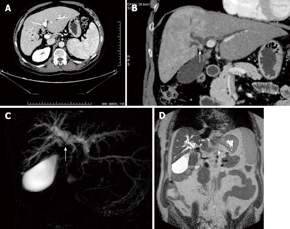
Figure 4 Periductal cholangiocarcinoma multidetector computed tomography and magnetic resonance cholangiopancreatography.
A: Multidetector computed tomography in the portal phase shows periductal cholangiocarcinoma (arrow) producing complete biliary obstruction and atrophy of the right liver; B: Coronal reconstruction shows to a better advantage the scirrhous infiltrating cholangiocarcinoma slightly hyperenhancing (arrow) and the dilated bile ducts upstream; C: Coronal thick-slab (echo spacing 8.3 ms, effective echo time 1000 ms, image matrix 512 x 512, FOV 350 mm) MRCP T2-W sequence in the same patient shows marked dilatation of intrahepatic bile ducts and a signal void (arrow) at the level of the hepatic convergence and common hepatic duct consistent with malignant obstruction by cholangiocarcinoma; D: Matrix (272 x 512, FOV 385 mm) T2-W sequence shows marked dilatation of the intrahepatic ducts and a narrow stenosis at the hepatic bifurcation (arrow).
- Citation: Valls C, Ruiz S, Martinez L, Leiva D. Radiological diagnosis and staging of hilar cholangiocarcinoma. World J Gastrointest Oncol 2013; 5(7): 115-126
- URL: https://www.wjgnet.com/1948-5204/full/v5/i7/115.htm
- DOI: https://dx.doi.org/10.4251/wjgo.v5.i7.115


