Copyright
©The Author(s) 2024.
World J Gastrointest Oncol. Feb 15, 2024; 16(2): 550-556
Published online Feb 15, 2024. doi: 10.4251/wjgo.v16.i2.550
Published online Feb 15, 2024. doi: 10.4251/wjgo.v16.i2.550
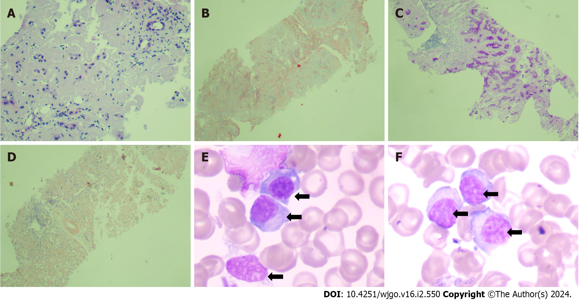
Figure 2 Liver pathology and bone marrow aspiration smear.
A: Hematoxylin-eosin staining (100 ×) showed deposition of pink fine-grained amorphous material in hepatic lobule and portal area, compression and deformation of liver plates, and atrophy of hepatocytes; B: Masson staining (40 ×) showed mild peri-sinusoidal fibrosis; C: Periodic acid-Schiff staining (40 ×) showed significant atrophy of liver cells with a few residual liver cells; D: Congo red staining (40 ×) indicated brick-red material deposition between liver cells; E and F: Bone marrow aspiration smear (100 ×) showed abnormal proliferation of plasma cell system. The cells are moderate-sized, the nucleus is large and eccentric, round or oval, the nuclear chromatin is fine, the cytoplasm is rich, blue, and foamy (black arrows).
- Citation: Zhang X, Tang F, Gao YY, Song DZ, Liang J. Hepatomegaly and jaundice as the presenting symptoms of systemic light-chain amyloidosis: A case report. World J Gastrointest Oncol 2024; 16(2): 550-556
- URL: https://www.wjgnet.com/1948-5204/full/v16/i2/550.htm
- DOI: https://dx.doi.org/10.4251/wjgo.v16.i2.550


