Copyright
©The Author(s) 2017.
World J Gastrointest Oncol. Sep 15, 2017; 9(9): 385-389
Published online Sep 15, 2017. doi: 10.4251/wjgo.v9.i9.385
Published online Sep 15, 2017. doi: 10.4251/wjgo.v9.i9.385
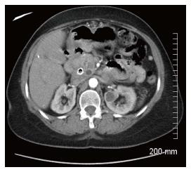
Figure 1 Computerized of the abdomen: Mass in head of pancreas, a dilated common bile duct with a metallic stent in place and a dilated pancreatic duct.
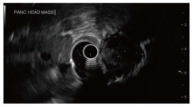
Figure 2 Endoscopic ultrasound showing a 2.
5 cm × 2.2 cm hypoechoic mass in the head of the pancreas causing extrahepatic biliary obstruction and pancreatic ductal dilation.
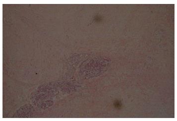
Figure 3 Histologic section (H and E) of the pancreatic head tumor.
Diffusely infiltrative tumor replacing parenchyma with focal remnants of normal appearing epithelial structures.
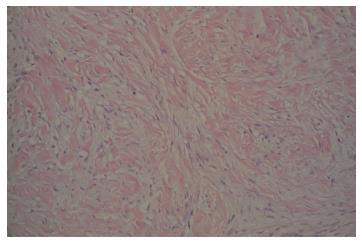
Figure 4 Typical DTF histology (H and E): Uniform sweeping fascicles of spindled myofibroblasts with low cellularity, no cellular atypia, minimal inflammation and scattered keloid-like collagen within a collagenous stroma.
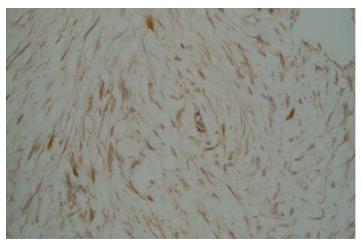
Figure 5 Beta-catenin immunohistochemistry: Positive, diffuse, plump myofibroblast nuclear staining, faint cytoplasmic staining characteristic of desmoid type fibromatosis.
- Citation: Jafri SF, Obaisi O, Vergara GG, Cates J, Singh J, Feeback J, Yandrapu H. Desmoid type fibromatosis: A case report with an unusual etiology. World J Gastrointest Oncol 2017; 9(9): 385-389
- URL: https://www.wjgnet.com/1948-5204/full/v9/i9/385.htm
- DOI: https://dx.doi.org/10.4251/wjgo.v9.i9.385


