Copyright
©The Author(s) 2016.
World J Gastrointest Oncol. Dec 15, 2016; 8(12): 835-839
Published online Dec 15, 2016. doi: 10.4251/wjgo.v8.i12.835
Published online Dec 15, 2016. doi: 10.4251/wjgo.v8.i12.835
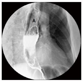
Figure 1 Barium esophagram showing an (A) obstructing intraluminal mass in the thoracic esophagus.
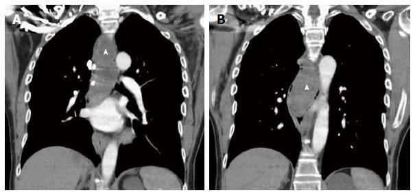
Figure 2 Computed tomographic scan of the chest (A) (B) showing a (arrowhead ) large mass traversing the length of the esophagus.
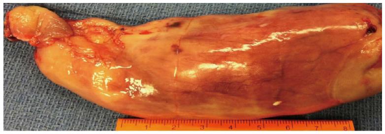
Figure 3 A macroscopic view of the resected giant fibrovascular polyp (13 cm × 6 cm × 2.
6 cm) with uniform surface and a large single stalk.
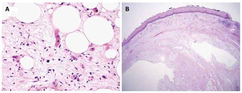
Figure 4 Histologically the polyp showed a central core of adipose and fibrovascular tissue surrounded by overlying squamous mucosa.
A: Hematoxylin and eosin stain × 40 identifying (arrowhead) striated muscle cells and adipose tissue within the core of the esophageal liposarcoma; B: The giant polyp is characterized by a central core of adipose and fibrovascular tissue surrounded by overlying squamous mucosa.
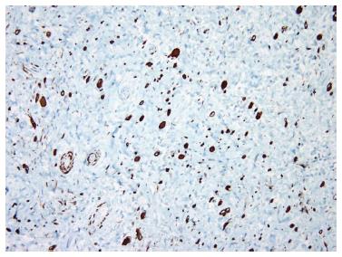
Figure 5 Immunohistology showed positive nuclear staining of lipoblasts with MDM2 confirming the diagnosis of liposarcoma.
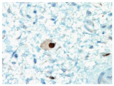
Figure 6 Cell with rhabdomyomatous differentiation were focally positive for myogenin.
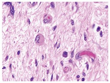
Figure 7 Rabdomyomatous differentiation is characterized by single and loose aggregates of large round cells with abundant eosinophilic cytoplasm.
- Citation: Valiuddin HM, Barbetta A, Mungo B, Montgomery EA, Molena D. Esophageal liposarcoma: Well-differentiated rhabdomyomatous type. World J Gastrointest Oncol 2016; 8(12): 835-839
- URL: https://www.wjgnet.com/1948-5204/full/v8/i12/835.htm
- DOI: https://dx.doi.org/10.4251/wjgo.v8.i12.835


