Copyright
©The Author(s) 2015.
World J Gastrointest Oncol. Nov 15, 2015; 7(11): 361-368
Published online Nov 15, 2015. doi: 10.4251/wjgo.v7.i11.361
Published online Nov 15, 2015. doi: 10.4251/wjgo.v7.i11.361
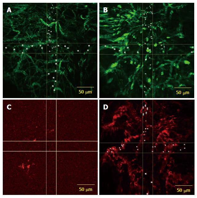
Figure 1 Confocal laser endomicroscopy.
A: CLE images with AF488 anti-CD31 antibodies expression on vascular network from both normal; B: Tumor rectal mucosa; C: CLE image showing low expression of the fluorescently labeled anti-CD105 antibodies in normal rectal mucosa; D: Image from the same patient showing microvessels in rectal adenocarcinoma visualized by using CD105 staining as a specific endothelial marker. CLE: Confocal laser endomicroscopy.
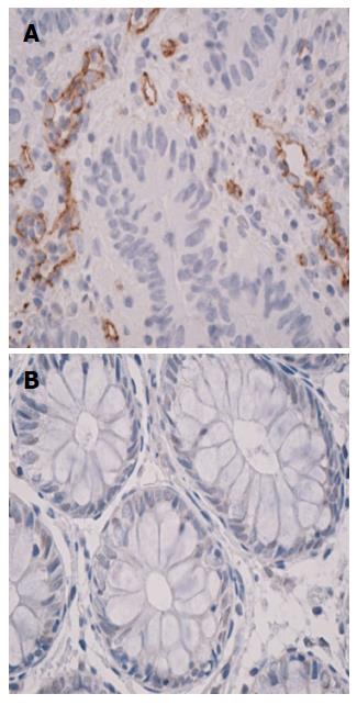
Figure 2 Immunohistochemistry on CD105 stained sequential sections from rectal cancer tissue samples (magnification 40 ×), CD105-positive vascular endothelial cells were clearly identified by their brown staining (A) and normal rectal mucosa displays the absence of endoglin expression (B).
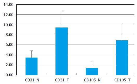
Figure 3 Graphic representation of vascular density (microvessel density) obtained from CD31-immunostained images and CD105-immunostained images of normal mucosa in comparison with tumor mucosa (vessels/mm2).
MVD: Microvessel density.
- Citation: Ciocâlteu A, Săftoiu A, Pirici D, Georgescu CV, Cârţână T, Gheonea DI, Gruionu LG, Cristea CG, Gruionu G. Tumor neoangiogenesis detection by confocal laser endomicroscopy and anti-CD105 antibody: Pilot study. World J Gastrointest Oncol 2015; 7(11): 361-368
- URL: https://www.wjgnet.com/1948-5204/full/v7/i11/361.htm
- DOI: https://dx.doi.org/10.4251/wjgo.v7.i11.361


