Copyright
©2013 Baishideng Publishing Group Co.
World J Gastrointest Oncol. Nov 15, 2013; 5(11): 198-203
Published online Nov 15, 2013. doi: 10.4251/wjgo.v5.i11.198
Published online Nov 15, 2013. doi: 10.4251/wjgo.v5.i11.198
Figure 1 Chest computed tomography-scan demonstrating a 7.
9 cm × 7.8 cm mass in the right upper lobe and right sided mediastinal and hilar lymphadenopathy.
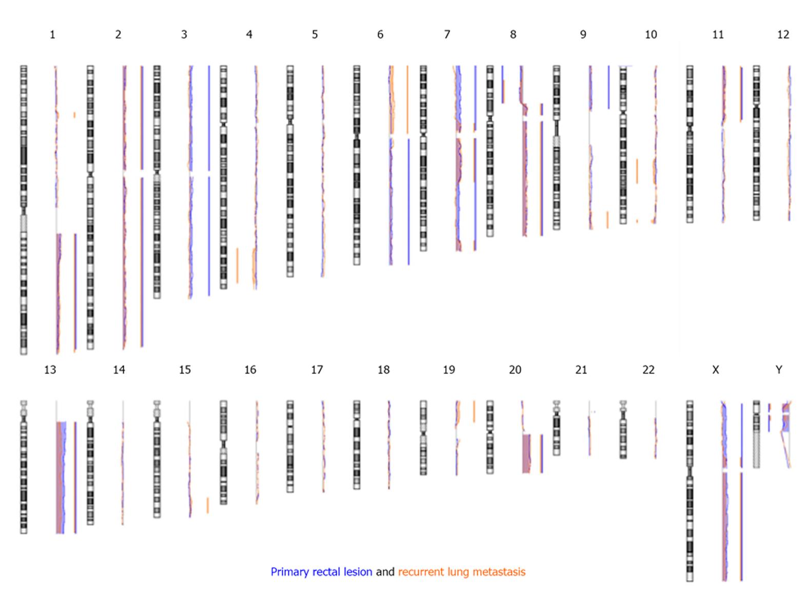
Figure 2 Common aberrations between the rectal tumor and the lung metastasis.
The abnormalities are summarized by the colored bars (blue for the colon tumor and orange for the lung metastasis). The bar is to the right of the tracing when there is DNA gain and to the left of the tracing when there is DNA loss. The length of the bar delineates the area of the chromosome involved.
- Citation: Rahma OE, Burotto M, Do Canto LM, Germanos AA, Haddad BR, Marshall JL. Striking similarities in genetic aberrations between a rectal tumor and its lung recurrence. World J Gastrointest Oncol 2013; 5(11): 198-203
- URL: https://www.wjgnet.com/1948-5204/full/v5/i11/198.htm
- DOI: https://dx.doi.org/10.4251/wjgo.v5.i11.198


