Copyright
©2012 Baishideng.
World J Gastrointest Oncol. Jun 15, 2012; 4(6): 152-155
Published online Jun 15, 2012. doi: 10.4251/wjgo.v4.i6.152
Published online Jun 15, 2012. doi: 10.4251/wjgo.v4.i6.152
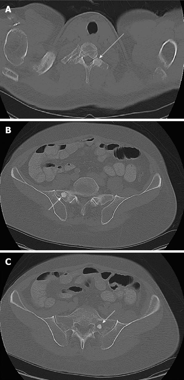
Figure 1 Axial computed tomography scan of the patient.
A: Computed tomography (CT) through the upper chest displayed using the bone window settings. There is a 2-cm well-defined sclerotic lesion within the left transverse process of the T1 vertebra (arrow); B: CT through the pelvis displayed using the bone window settings. There is a 1.5-cm, well defined, sclerotic lesion (arrow) in the right sacrum adjacent to the sacroiliac joint; C: CT through the pelvis displayed using the bone window settings. There is a 1.5-cm, well defined, sclerotic lesion (arrow) in the left sacrum.
- Citation: Ghetie C, Cornfeld D, Ramfidis VS, Syrigos KN, Saif MW. Bone lesions in recurrent glucagonoma: A case report and review of literature. World J Gastrointest Oncol 2012; 4(6): 152-155
- URL: https://www.wjgnet.com/1948-5204/full/v4/i6/152.htm
- DOI: https://dx.doi.org/10.4251/wjgo.v4.i6.152


