Copyright
©2013 Baishideng Publishing Group Co.
World J Gastrointest Oncol. Nov 15, 2012; 4(11): 216-222
Published online Nov 15, 2012. doi: 10.4251/wjgo.v4.i11.216
Published online Nov 15, 2012. doi: 10.4251/wjgo.v4.i11.216
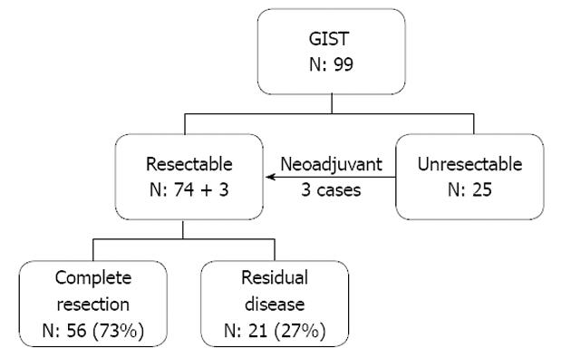
Figure 1 Categorization of 99 cases of gastrointestinal intestinal tumor according to their resectability.
GIST: Gastrointestinal stromal tumor; N: Number.
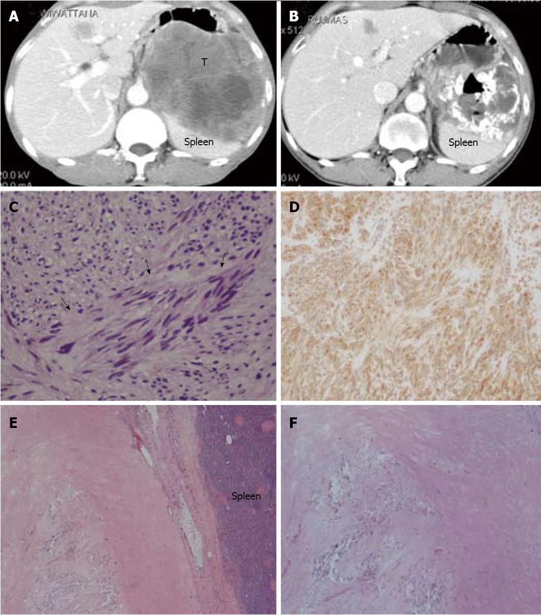
Figure 2 Abdominal computerized tomography and histopathological pictures of a 68-year-old male patient who presented with abdominal mass.
A: Computerized tomography (CT) shows a large enhancing solid mass (T), measuring 11.2 cm × 11.9 cm × 10.7 cm, occupying the left upper quadrant between the stomach and the spleen. A liver nodule is also visible in segment IV; B: Forty months following the beginning of imatinib therapy, a follow-up CT showed partial tumor response; C, D: Image-guided tissue biopsy revealed a spindle cell tumor (arrows) that marked CD117. The mitotic cell count was 2 cells/50 high power fields; E, F: Following an en bloc resection including a total removal of the stomach together with the spleen and a wedge resection of hepatic metastasis, the pathological tissue showed only a stromal hyalinization and dystrophic calcification with a scanty number of differentiated spindle cells that marked S-100, but not CD117.
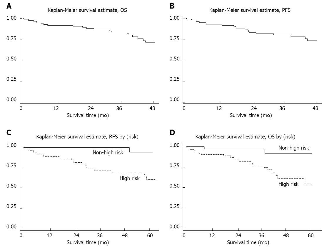
Figure 3 Kaplan-Meier survival probability curves.
Kaplan-Meier survival probability curves showing overall survival (OS) (A) progress free survival (PFS) (B), significant difference in the recurrent free survival (RFS) after surgery in primary resectable cases, comparing between the cases in the high risk group according to the National Institute of Health risk categorization, and the cases in the other risk groups (C) and significant difference in the OS (D).
- Citation: Pornsuksiri K, Chewatanakornkul S, Kanngurn S, Maneechay W, Chaiyapan W, Sangkhathat S. Clinical outcomes of gastrointestinal stromal tumor in southern Thailand. World J Gastrointest Oncol 2012; 4(11): 216-222
- URL: https://www.wjgnet.com/1948-5204/full/v4/i11/216.htm
- DOI: https://dx.doi.org/10.4251/wjgo.v4.i11.216


