Copyright
©2011 Baishideng Publishing Group Co.
World J Gastrointest Oncol. Jul 15, 2011; 3(7): 111-115
Published online Jul 15, 2011. doi: 10.4251/wjgo.v3.i7.111
Published online Jul 15, 2011. doi: 10.4251/wjgo.v3.i7.111
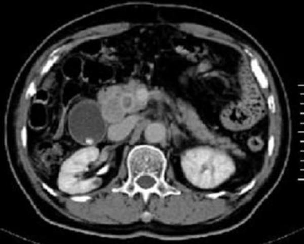
Figure 1 Computed tomography showing a pancreas head tumor, measuring 30 mm × 20 mm, and a dilated common bile duct.
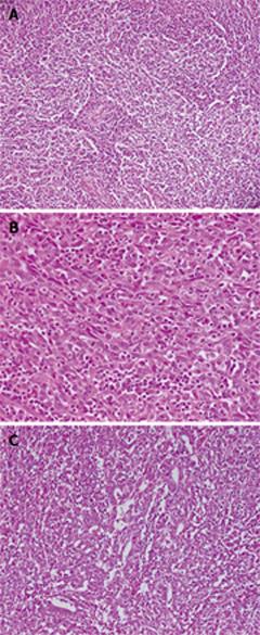
Figure 2 Histopathological findings.
A: Nests of undifferentiated cells in a background of dense lymphoplasmacytic infiltration (hematoxylin and eosion stain, × 100); B: Undifferentiated tumor cells with irregular large vesicular nuclei with nucleoli, and densely infiltrated lymphocytes and plasma cells that appear mature (hematoxylin and eosion stain, × 200); C: Focal glandular differentiation is present (hematoxylin and eosion stain, × 100).
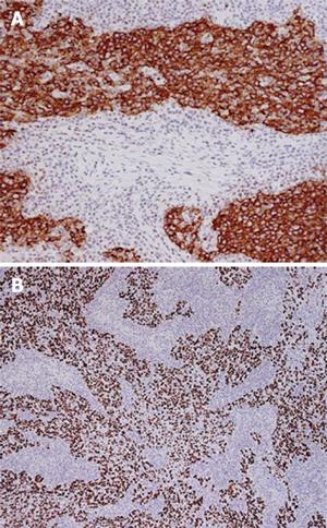
Figure 3 Immunohistochemical findings.
A: CA19-9 expression in tumor cells (× 100); B: Overexpression of p53 protein in tumor cells (× 40).
- Citation: Ishida M, Mori T, Shiomi H, Naka S, Tsujikawa T, Andoh A, Saito Y, Kurumi Y, Kojima F, Hotta M, Tani T, Fujiyama Y, Okabe H. Non-Epstein-Barr virus associated lymphoepithelioma-like carcinoma of the inferior common bile duct. World J Gastrointest Oncol 2011; 3(7): 111-115
- URL: https://www.wjgnet.com/1948-5204/full/v3/i7/111.htm
- DOI: https://dx.doi.org/10.4251/wjgo.v3.i7.111


