Copyright
©2010 Baishideng.
World J Gastrointest Oncol. May 15, 2010; 2(5): 222-228
Published online May 15, 2010. doi: 10.4251/wjgo.v2.i5.222
Published online May 15, 2010. doi: 10.4251/wjgo.v2.i5.222
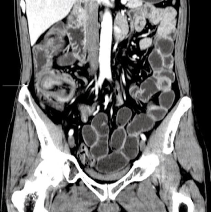
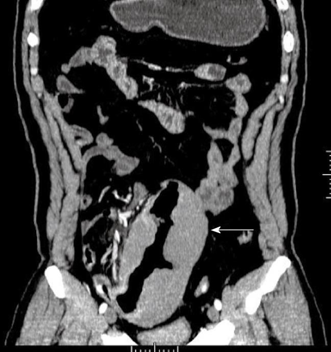
Figure 1 Adenoma of the ileum in a 54-year-old man who presented with a 1-year history of abdominal fullness and pain with hematochezia.
Contrast-enhanced computed tomography (CT) scan shows a homogenous, moderate enhanced mass and ileocolic intussusceptions with thickening of the terminal ileum (white arrow).
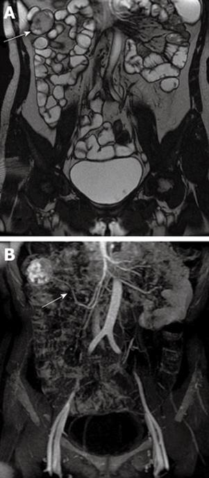
Figure 2 Gastrointestinal stromal tumor of the ileum in a 52-year-old woman who presented with persistent abdominal pain and hematochezia.
A: Gadolinium-enhanced T2-weighted image shows a well-circumscribed mass with heterogeneous intermediate signal intensity (white arrow); B: MIP image shows the tumor was supplied by the superior mesenteric artery (white arrow). MIP: Maximum intensity projection.
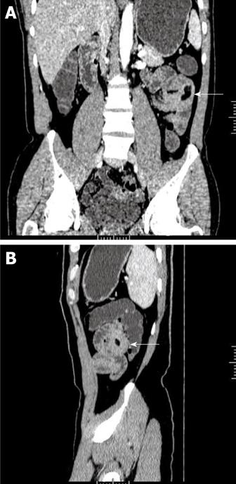
Figure 3 Lipoma of the jejunum in a 56-year-old man who presented with a 3-year history of hematochezia.
A: Image (Coronal MPR) of contrast-enhanced CT scan shows a homogeneous fat-tissue density mass in jejunal smooth muscle (white arrow); B: Image (Sagittal MPR) shows obviously thickened intestinal wall (white arrow). MPR: Multiplanar reconstruction.
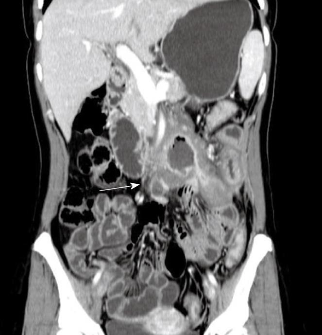
Figure 4 Adenocarcinoma of the jejunum in a 38-year-old woman who presented with weight loss, abdominal pain and an abdominal mass.
Contrast-enhanced CT scan shows a large, moderate enhanced soft tissue mass without a clear edge and infiltration of mesenteric vessels (white arrow).
Figure 5 Diffuse large B-cell lymphoma of the ileum in a 46-year-old man who presented with a 2-year history of abdominal pain and intermittent diarrhea and was previously suspected as having Crohn’s Disease.
Contrast-enhanced CT scan shows diffuse, homogeneous wall thickening of the ileum, aneurysmal dilatation of the lumen and no bowel obstruction (white arrow).
- Citation: Miao F, Wang ML, Tang YH. New progress in CT and MRI examination and diagnosis of small intestinal tumors. World J Gastrointest Oncol 2010; 2(5): 222-228
- URL: https://www.wjgnet.com/1948-5204/full/v2/i5/222.htm
- DOI: https://dx.doi.org/10.4251/wjgo.v2.i5.222


