Copyright
©2010 Baishideng Publishing Group Co.
World J Gastrointest Oncol. Oct 15, 2010; 2(10): 390-394
Published online Oct 15, 2010. doi: 10.4251/wjgo.v2.i10.390
Published online Oct 15, 2010. doi: 10.4251/wjgo.v2.i10.390
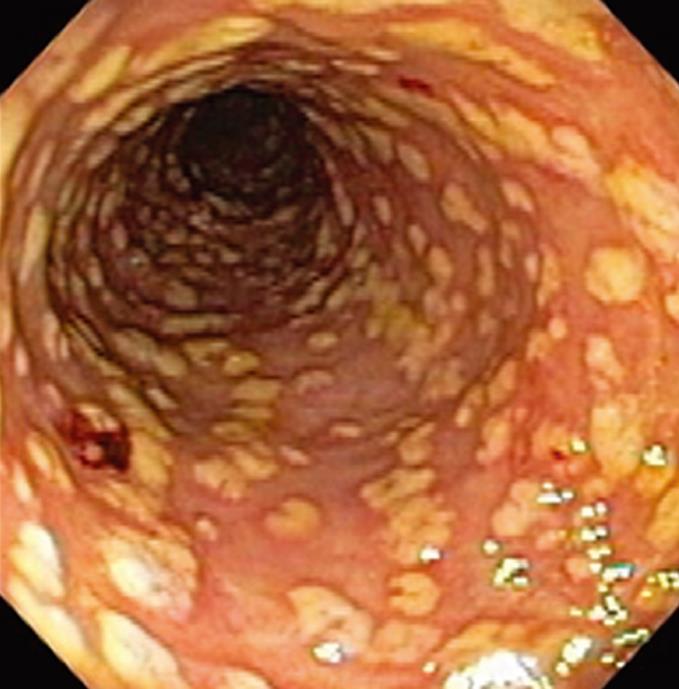
Figure 1 Endoscopic appearance of the sigmoid colon in case 1 showing diffuse pseudomembranes with hemorrhagic.
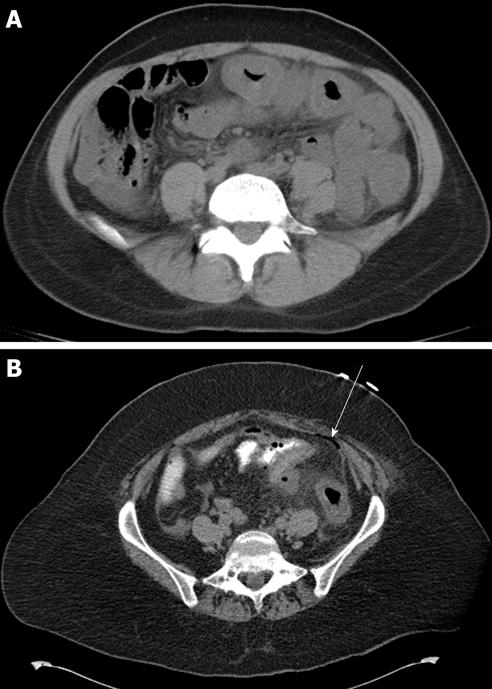
Figure 2 Computed tomographic scan of the abdomen in case 3 (A) and case 4 (B).
A: Thickening of the walls of the transverse and descending colon; B: Thickening of the walls of the descending colon and sigmoid with pneumatosis and free peritoneal air (white arrow).
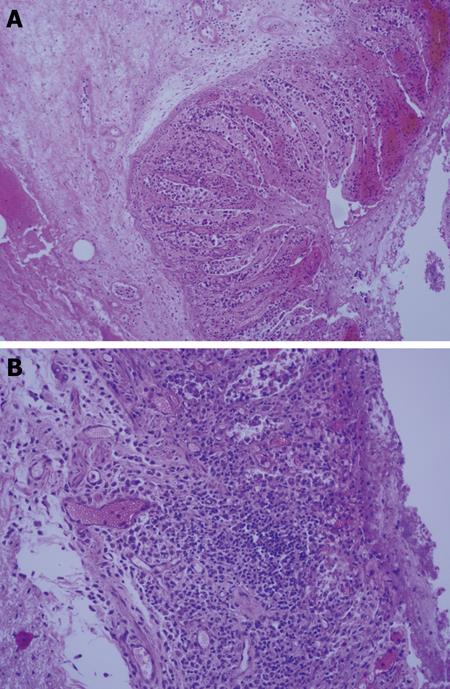
Figure 3 Microscopic appearance of a resected colonic segment in case 4.
A: Mucosal and submucosal edema, hemorrhage, acute inflammation and necrosis (H&E, 200 × magnification); B: Transmural necrosis (H&E, 200 × magnification).
- Citation: Carrion AF, Hosein PJ, Cooper EM, Lopes G, Pelaez L, Rocha-Lima CM. Severe colitis associated with docetaxel use: A report of four cases. World J Gastrointest Oncol 2010; 2(10): 390-394
- URL: https://www.wjgnet.com/1948-5204/full/v2/i10/390.htm
- DOI: https://dx.doi.org/10.4251/wjgo.v2.i10.390


