
















Copyright: ©Author(s) 2026.
World J Gastrointest Oncol. Nov 15, 2026; 18(11): 124427
Published online Nov 15, 2026. doi: 10.4251/wjgo.124427
Published online Nov 15, 2026. doi: 10.4251/wjgo.124427
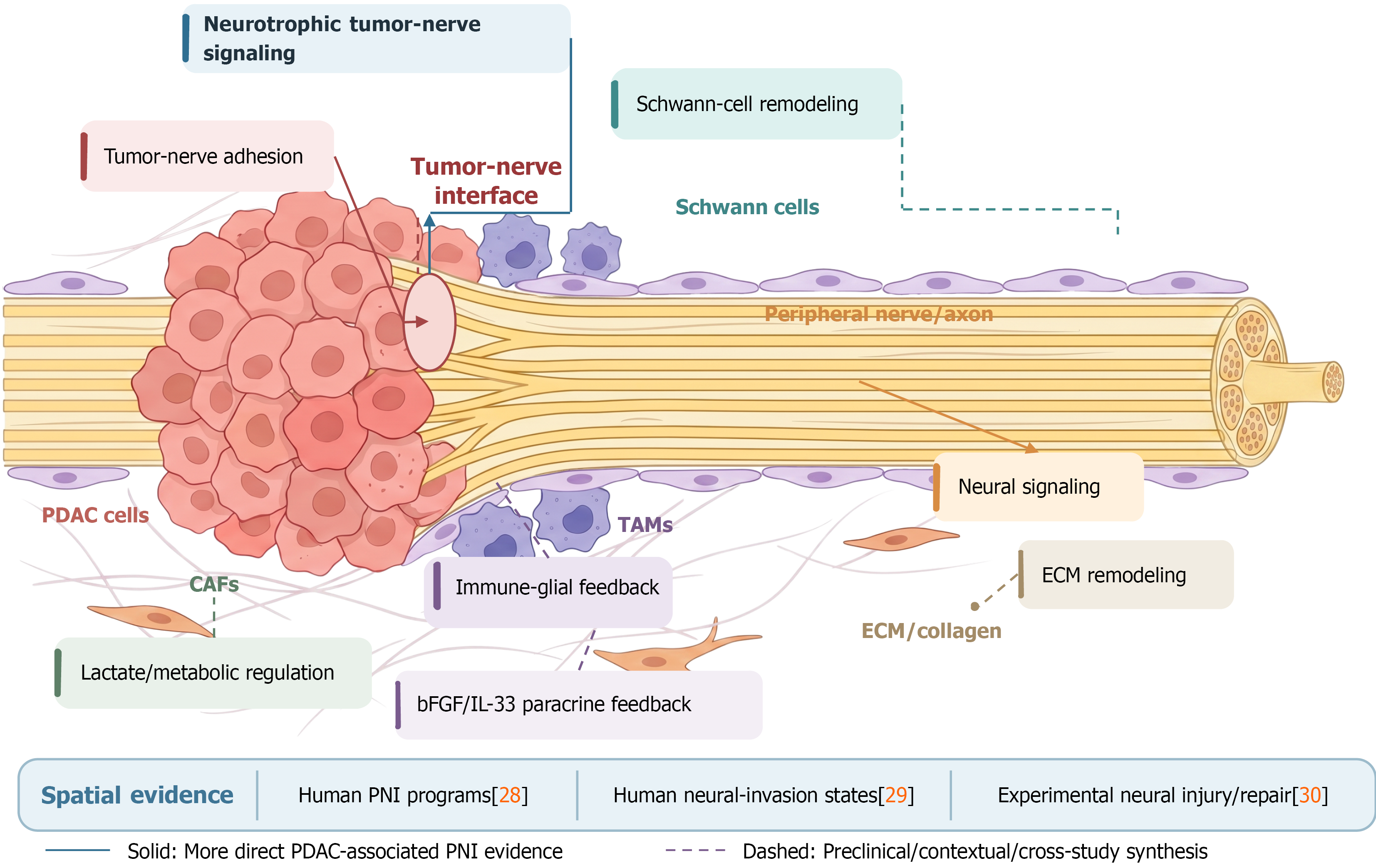
Figure 1 Neural-invasion niche and molecular mechanisms in pancreatic ductal adenocarcinoma.
Some graphic elements in the images come from the BioRender standard materials/icons library and were used in accordance with BioRender’s license. This figure illustrates the neural-immune-stromal-metabolic niche associated with perineural invasion (PNI) in pancreatic ductal adenocarcinoma (PDAC). The central anatomical interface consists of PDAC cells approaching and interacting with a peripheral nerve, together with Schwann cells, TAMs, cancer-associated fibroblasts, and extracellular matrix. Neurotrophic tumor–nerve signaling, including nerve growth factor/tropomyosin receptor kinase A - and glial cell line-derived neurotrophic factor/rearranged during transfection-related pathways, may promote tumor-nerve approximation and invasive behavior. Tumor-nerve adhesion further stabilizes contact at the interface. Schwann-cell remodeling may provide a permissive neural microenvironment for tumor-cell migration. Immune–glial signaling, including the TAMs-Schwann-cell basic fibroblast growth factor/interleukin-33 paracrine circuit, may reinforce local neural and immune remodeling. Extracellular matrix remodeling may provide a migration-permissive structural environment, whereas CAF-associated lactate metabolism and related RNA/epigenetic programs may further modify PNI-associated tumor behavior. Neural signaling represents additional communication between the nerve compartment and the surrounding TME. Arrowheads indicate the direction of the schematic relationship toward the relevant cellular or anatomical compartment. Solid connectors indicate relationships more directly supported by PDAC/PNI evidence; dashed connectors indicate predominantly preclinical, contextual, or integrative relationships. The spatial-evidence panel distinguishes direct human PNI spatial evidence[28], human neural-invasion-associated cellular states[29], and predominantly experimental neural injury/repair evidence[30]. These evidence categories indicate differences in the source and maturity of evidence rather than therapeutic efficacy. PDAC: Pancreatic ductal adenocarcinoma; CAFs: Cancer-associated fibroblasts; bFGF: Basic fibroblast growth factor; TAMs: Tumor-associated macrophages; PNI: Perineural invasion; ECM: Extracellular matrix; IL-33: Interleukin-33.
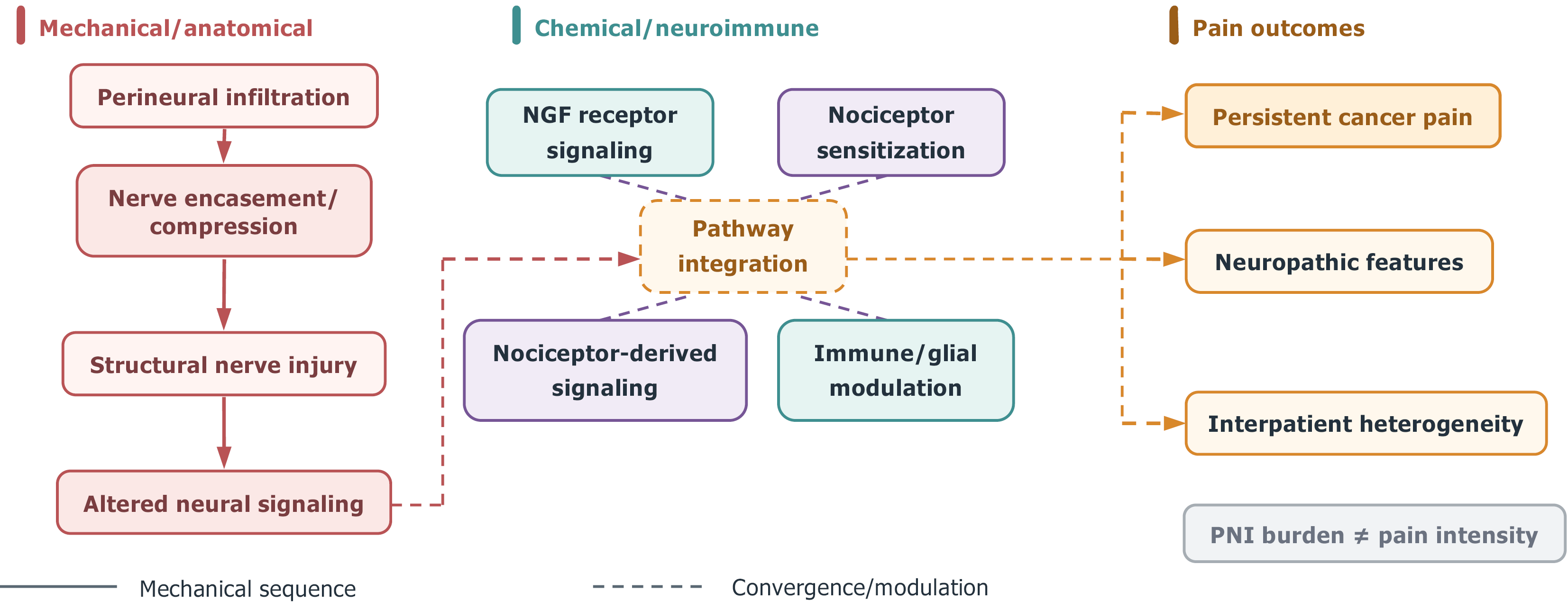
Figure 2 Pain generation and sensitization associated with perineural invasion in pancreatic ductal adenocarcinoma.
This figure summarizes two overlapping pathways involved in pain associated with perineural invasion in pancreatic ductal adenocarcinoma. On the left is the mechanical and anatomical pathway. After tumor cells infiltrate the perineural space, they may further encase or compress nerves, causing structural nerve damage and altering local neural signaling. These anatomical changes may constitute the structural basis for the characteristics of persistent cancer pain and neuropathic pain. The middle section depicts the chemical and neuroimmune pathway. Nerve growth factor receptor signaling may enhance the responsiveness of sensory neurons and promote sensitization of nociceptors; signals from nociceptors, along with modulation by immune and glial cells, may further increase local neural sensitivity. The right-hand section illustrates clinical pain outcomes that may arise from the integration of mechanical/anatomical injury and chemical/neuroimmune sensitization, including persistent cancer pain, neuropathic features, and interpatient heterogeneity. Solid lines show the mechanical/anatomical sequence from perineural infiltration to nerve sheath compression, structural nerve damage, and changes in nerve signaling. Dashed lines represent the convergence or regulation between altered nerve signals, nerve growth factor-related sensitization, nociceptor signaling, immune and glial modulation, and pain outcomes. The statement “PNI burden ≠ pain intensity” in the figure emphasizes that the anatomical burden of perineural invasion does not directly determine pain intensity. Pain in different patients may result from a combination of structural nerve damage and chemical/neuroimmune sensitization to varying degrees. PNI: Perineural invasion; NGF: Nerve growth factor.
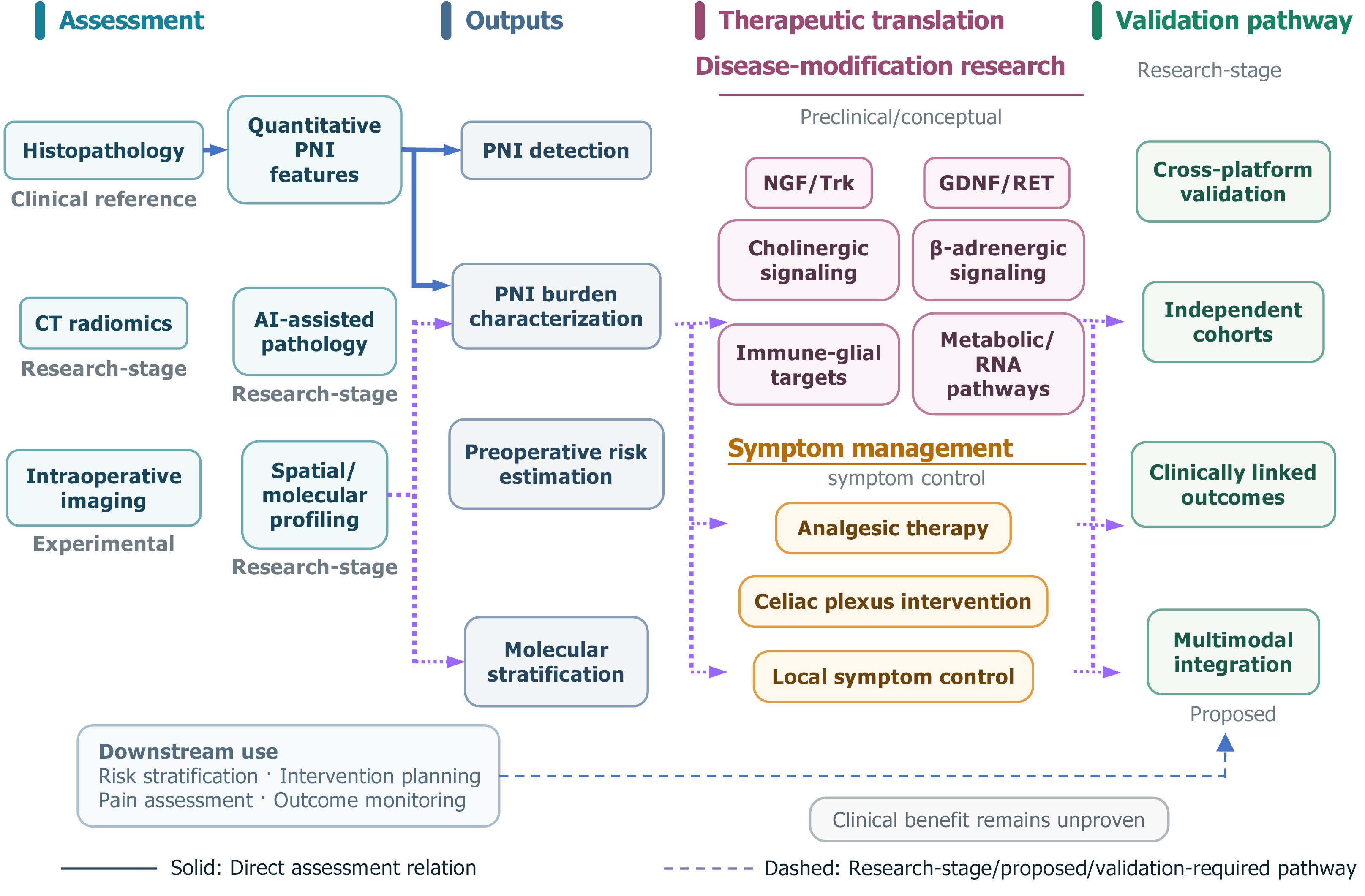
Figure 3 Diagnostic assessment, therapeutic translation, and validation road map for perineural invasion in pancreatic ductal adeno
- Citation: Hu R, Zhang HC, Zhang XF. Perineural invasion in pancreatic ductal adenocarcinoma: Molecular mechanisms and clinical significance. World J Gastrointest Oncol 2026; 18(11): 124427
- URL: https://www.wjgnet.com/1948-5204/full/v18/i11/124427.htm
- DOI: https://dx.doi.org/10.4251/wjgo.124427