










BPG is committed to discovery and dissemination of knowledge
Minireviews
Copyright: ©Author(s) 2026.
World J Gastrointest Oncol. Nov 15, 2026; 18(11): 124427
Published online Nov 15, 2026. doi: 10.4251/wjgo.124427
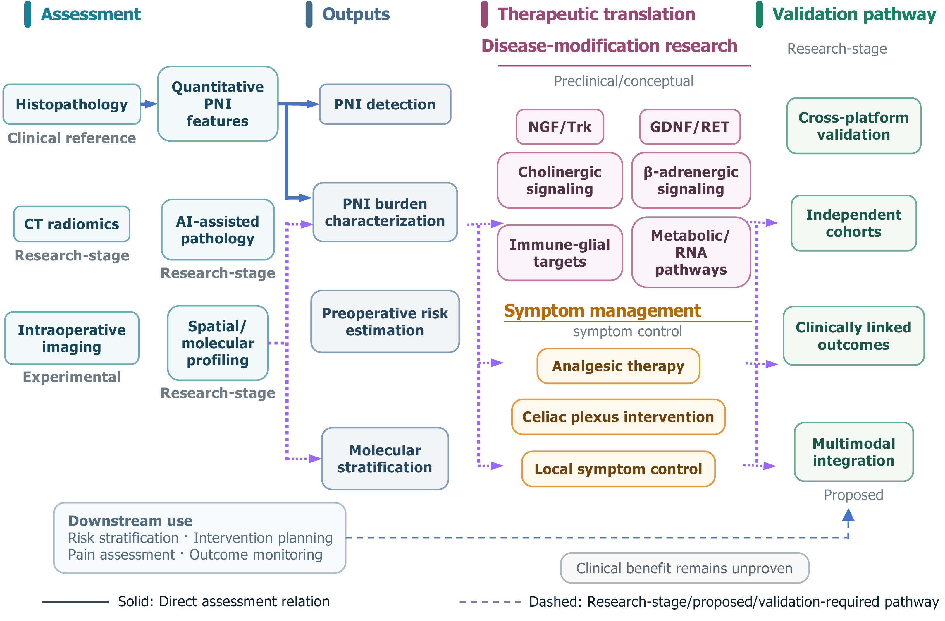
Figure 3
Figure 3 Diagnostic assessment, therapeutic translation, and validation road map for perineural invasion in pancreatic ductal adenocarcinoma. This figure summarizes the assessment methods of perineural invasion (PNI), the information obtained from these assessments, biological/molecular stratifications still at the research stage, therapeutic translation, and future validation requirements. The left section pertains to clinical diagnosis and post-assessment risk evaluation. Histopathology remains the clinical reference standard for PNI diagnosis, and quantitative features of PNI can further describe its detection and invasion burden. Computed tomography radiomics and artificial intelligence-assisted pathology are still in the research stage, while intraoperative imaging is mainly experimental. The information obtained from assessments includes PNI detection, characterization of PNI burden, preoperative risk evaluation, and molecular stratification, with the latter two not yet established as routine clinical tools. It is important to stress that spatial and molecular analysis is still a research-stage method for studying biological features and molecular layering, and it should not be seen as a routine tool for preoperative PNI risk assessment or treatment decisions. The third section concerns therapeutic translation, divided into disease-modification research and symptom management. Candidate molecular pathways mainly provide preclinical or conceptual guidance for therapeutic targets. Pain management, celiac plexus interventions, and local symptom control fall under symptom-oriented treatments. The fourth section outlines future validation pathways, including cross-platform validation, independent cohorts, correlation with clinical outcomes, and multimodal integration. Solid lines indicate relatively direct relationships within the current assessment framework, while dashed lines represent research-stage, proposed, or still-to-be-validated pathways, which do not imply established causal relationships or routine clinical procedures. The blue dashed line at the bottom of the figure connects potential downstream applications, including risk stratification, intervention planning, pain assessment, and outcome monitoring; however, their clinical application still requires further validation. Currently, there is no evidence showing that modifying PNI results in definite clinical benefits. PNI: Perineural invasion; CT: Computed tomography; AI: Artificial intelligence; NGF: Nerve growth factor; Trk: Tropomyosin receptor kinase; GDNF: Glial cell line-derived neurotrophic factor; RET: Rearranged during transfection.


Write to the Help Desk