
















Copyright: ©Author(s) 2026.
World J Gastrointest Oncol. Nov 15, 2026; 18(11): 123631
Published online Nov 15, 2026. doi: 10.4251/wjgo.123631
Published online Nov 15, 2026. doi: 10.4251/wjgo.123631
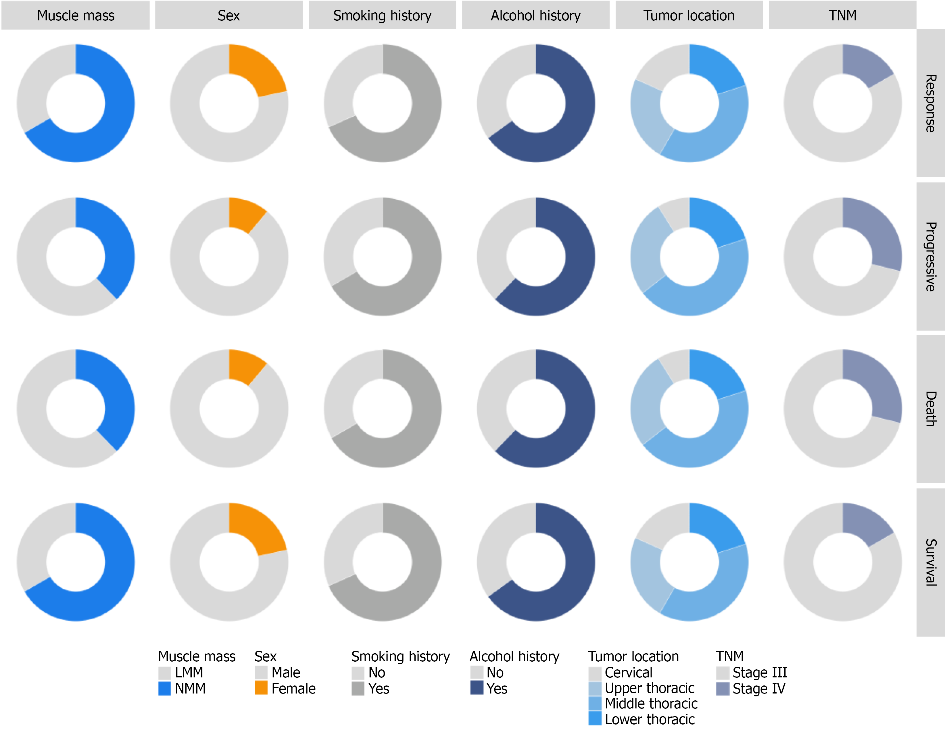
Figure 1 Clinical characteristics by treatment response and survival status.
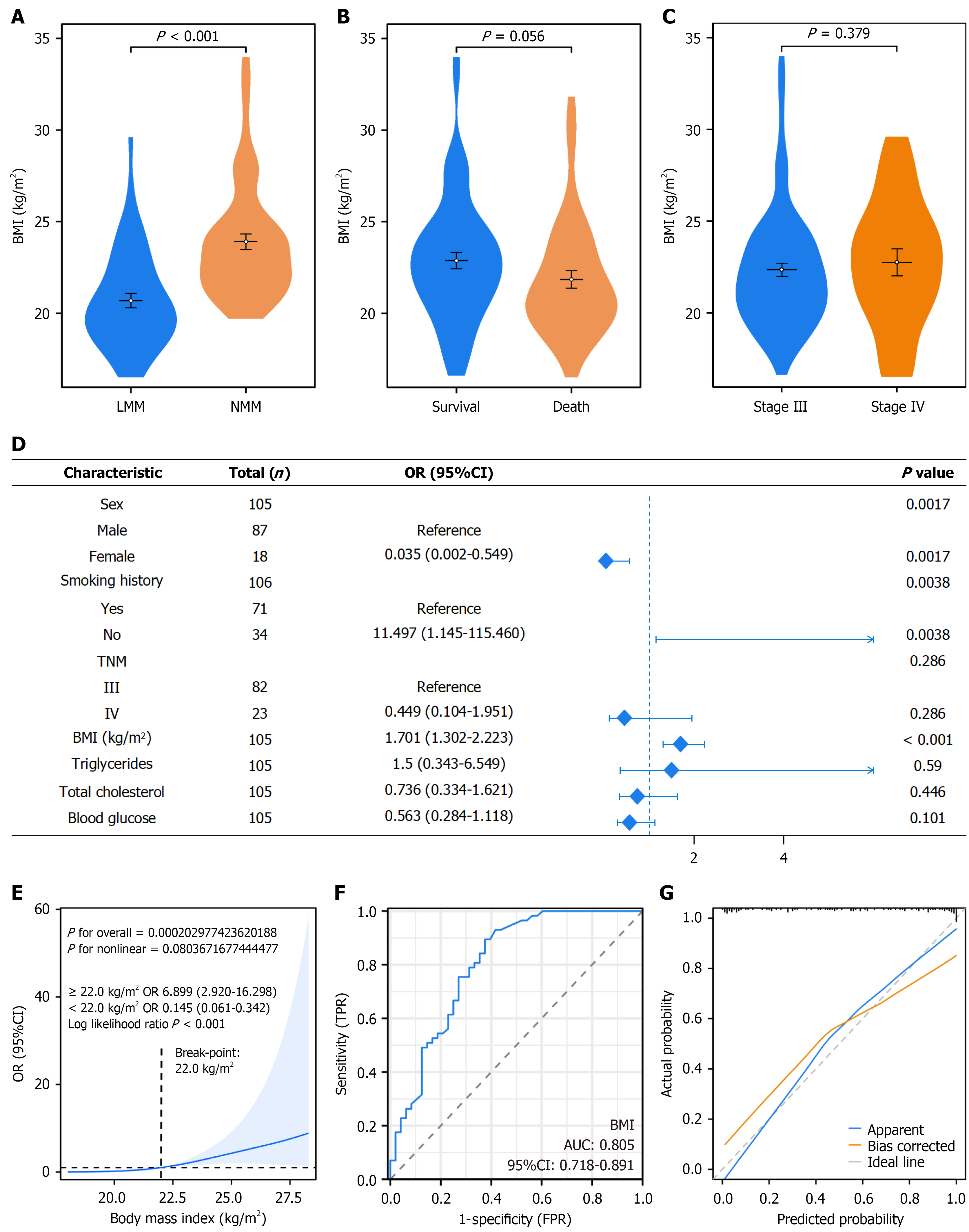
Figure 2 Low muscle mass and associated factors in esophageal cancer patients.
A-C: Present violin plots of body mass index (BMI) distribution across different clinical subgroups; D: A forest plot of multivariable logistic regression analysis for factors associated with low muscle mass (LMM); E: Depicts the nonlinear association between BMI and LMM using restricted cubic spline analysis; F and G: Display the receiver operating characteristic curve and calibration curve of the LMM prediction model, respectively. OR: Odds ratio; CI: Confidence interval; BMI: Body mass index; TNM: Tumor-node-metastasis; AUC: Area under the curve; FPR: False-positive rate; TPR: True-positive rate.
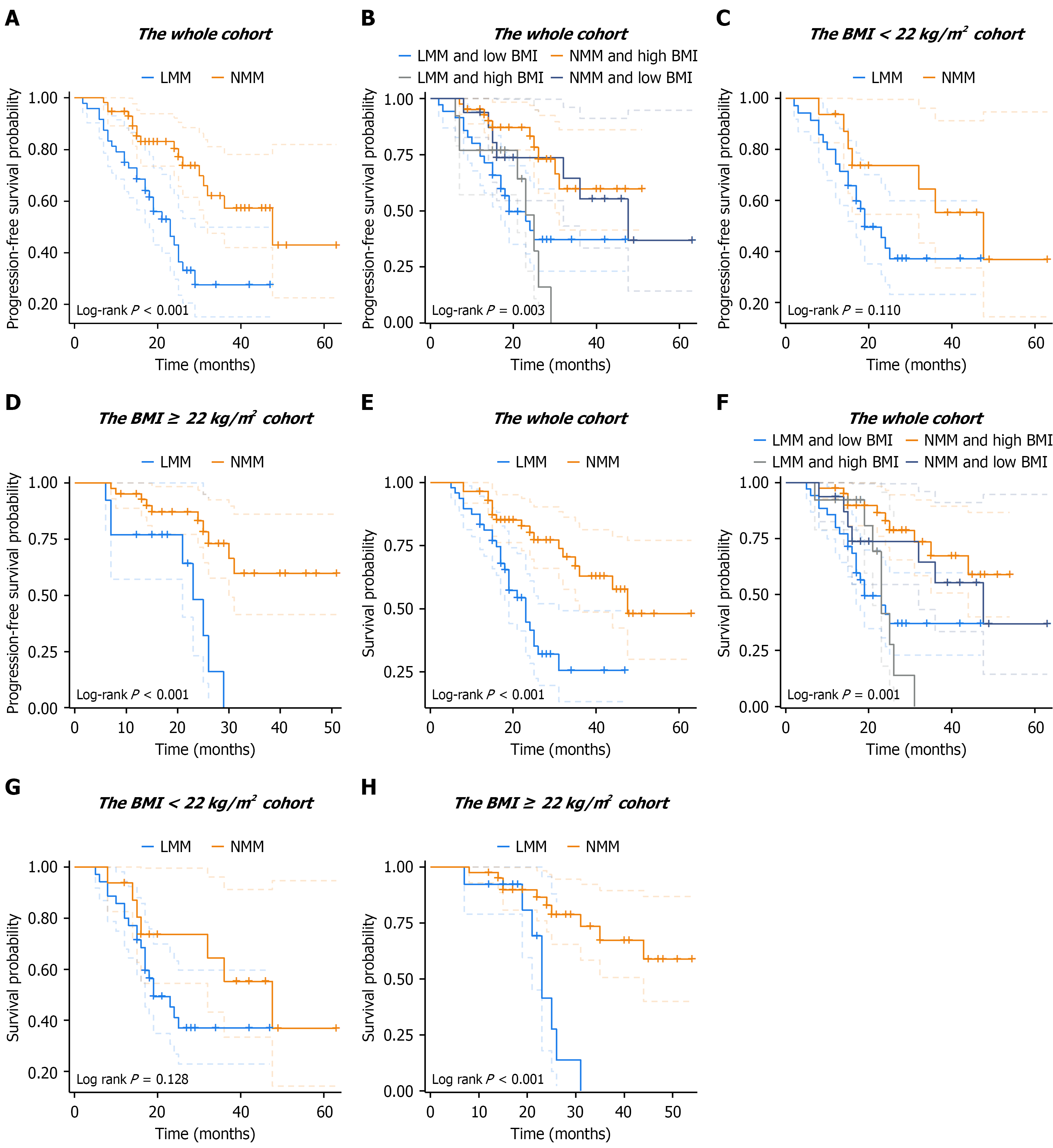
Figure 3 Survival analysis stratified by LMM and body mass index status in esophageal cancer patients.
A-D: Present progression-free survival (PFS); E-H: Present overall survival (OS). A and E: Comparison of PFS and OS between sarcopenic and non-sarcopenic patients in the entire cohort; B and F: Combined stratification by low muscle mass (LMM) and body mass index (BMI) status (four subgroups: LMM + low BMI, normal muscle mass + high BMI, LMM + high BMI, normal muscle mass + low BMI) for PFS and OS in the entire cohort; C and G: Stratified analyses of PFS and OS by LMM status in patients with BMI < 22 kg/m2; D and H: Stratified analyses of PFS and OS by LMM status in patients with BMI ≥ 22 kg/m2. NMM: Normal muscle mass; LMM: Low muscle mass; BMI: Body mass index.
- Citation: Xiao L, Li LJ, Lyu JH, Zhang SC, Liang L. Combined low muscle mass and body mass index for prognosis in esophageal squamous cell carcinoma patients receiving immunochemoradiotherapy. World J Gastrointest Oncol 2026; 18(11): 123631
- URL: https://www.wjgnet.com/1948-5204/full/v18/i11/123631.htm
- DOI: https://dx.doi.org/10.4251/wjgo.123631