
















Copyright: ©Author(s) 2026.
World J Gastrointest Oncol. Nov 15, 2026; 18(11): 123321
Published online Nov 15, 2026. doi: 10.4251/wjgo.123321
Published online Nov 15, 2026. doi: 10.4251/wjgo.123321
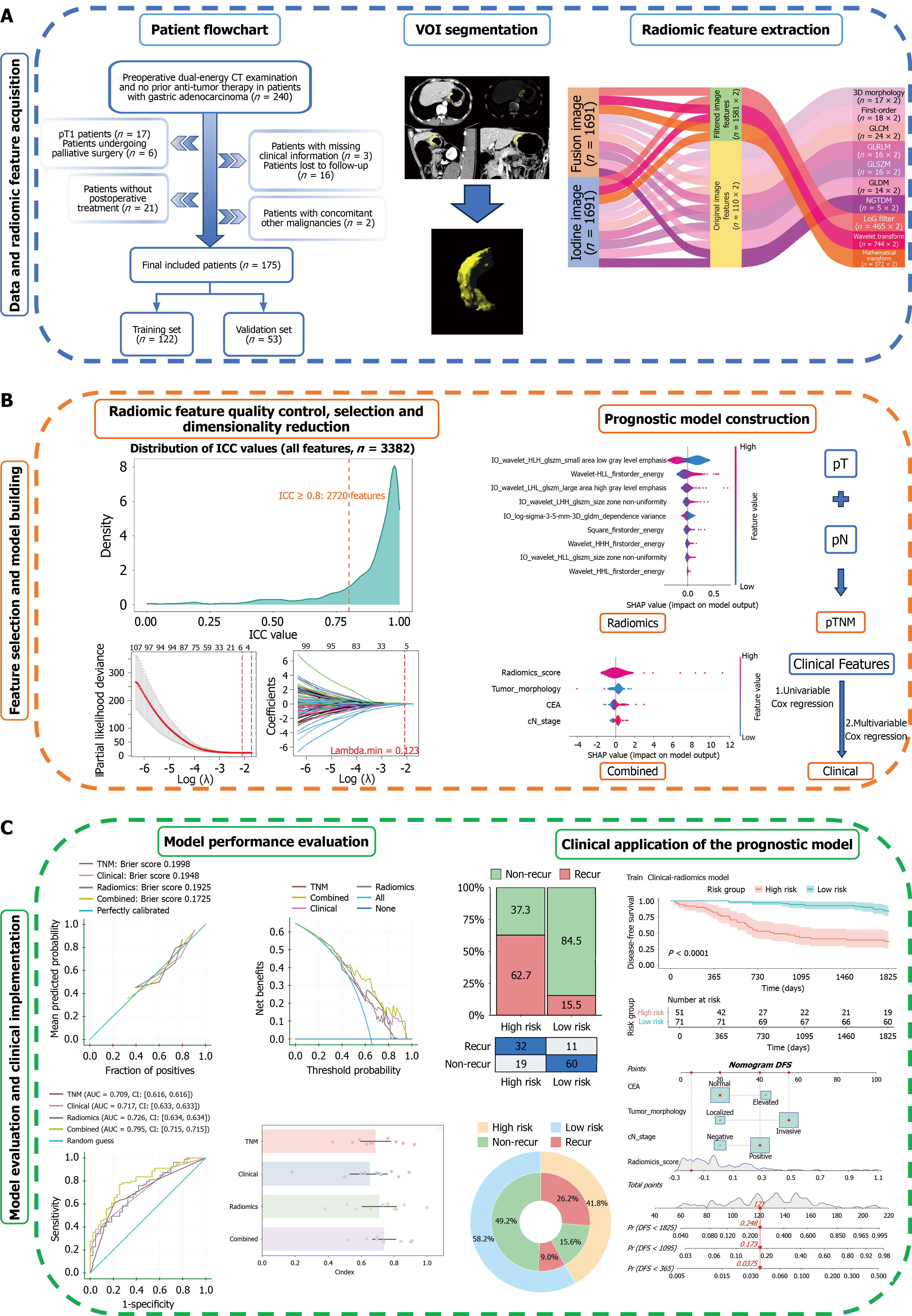
Figure 1 Study flowchart.
A: Acquisition of data and radiomic features: Including patient enrollment, image processing and volume of interest segmentation, and radiomic feature extraction; B: Screening of radiomic and clinical features, and construction of prognostic models; C: Performance evaluation and clinical application of the prognostic model. CT: Computed tomography; VOI: Volume of interest; ICC: Intraclass correlation coefficient; pTNM: Pathological tumor-node-metastasis; CEA: Carcinoembryonic antigen; AUC: Area under the curve; DFS: Disease-free survival.
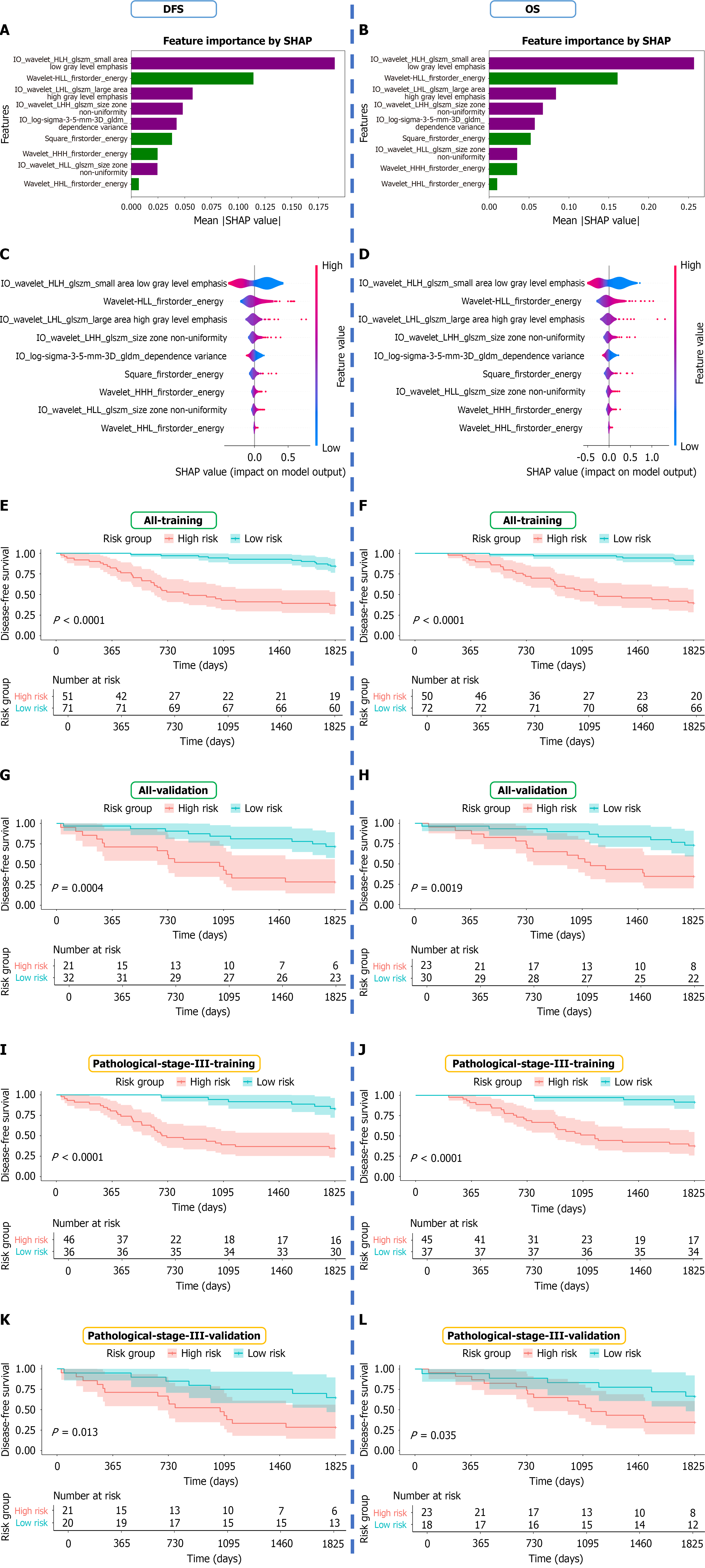
Figure 2 Feature contribution and Kaplan-Meier risk stratification of prognostic models.
A and B: Overall contribution of radiomic features to disease-free survival/overall survival prediction quantified by mean absolute SHapley Additive exPlanations values; C and D: Magnitude and direction of feature impact on model output visualized via SHapley Additive exPlanations summary plots; E-H: Kaplan-Meier curves of high- and low-risk subgroups among all patients in the training and validation sets; I-L: Kaplan-Meier curves of high- and low-risk subgroups among pathological stage III patients in the training and validation sets. DFS: Disease-free survival; OS: Overall survival.
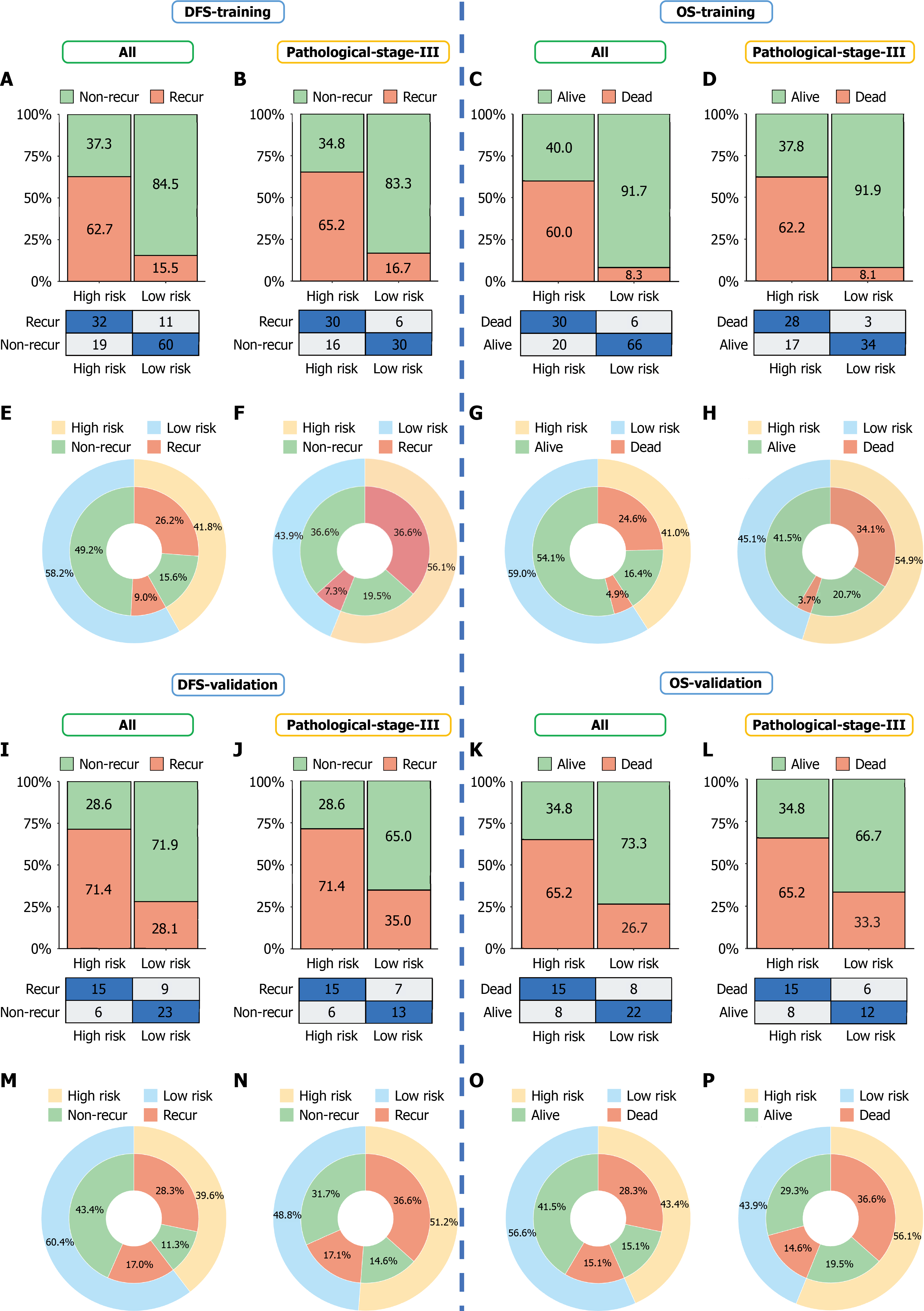
Figure 3 Confusion matrices and concentric plots showing the classification performance and clinical applicability of the combined model.
A-H: Confusion matrices and concentric plots for disease-free survival and overall survival prediction in all patients and patients with pathological stage III disease in the training set; I-P: Corresponding analyses in the validation set. DFS: Disease-free survival; OS: Overall survival; AIC: Akaike information criterion; TNM: Tumor-node-metastasis; AUC: Area under the curve; CI: Confidence interval.
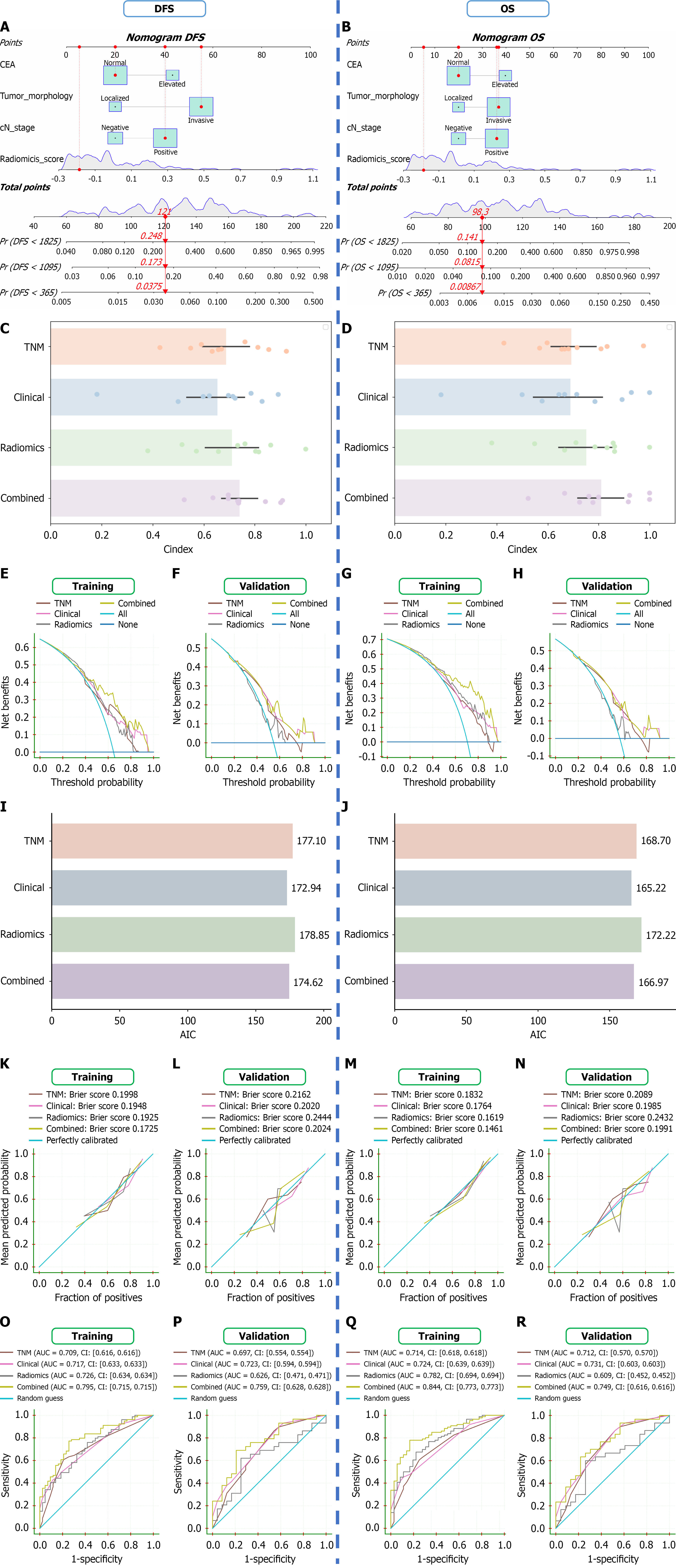
Figure 4 Nomograms and comprehensive model performance evaluation.
A and B: A patient with pT4N3M0 staging in the validation cohort was stratified into the low-risk group by the combined model; the nomogram indicated that the probabilities of the patient having disease-free survival and overall survival of less than 5 years were 24.8% and 14.1%, respectively. Ultimately, the patient remained alive at 5 years postoperatively without recurrence or metastasis; C and D: C-index comparison among models via 10-fold cross-validation in the training cohort; E-H: Decision curve analysis in the training and validation sets; I and J: Akaike information criterion values of each prognostic model in the validation set; K-N: Calibration curves in the training and validation sets; O-R: Receiver operating characteristic curves in the training and validation sets. DFS: Disease-free survival; OS: Overall survival; AIC: Akaike information criterion; TNM: Tumor-node-metastasis; AUC: Area under the curve; CI: Confidence interval.
- Citation: Liu A, Li M, You Y, Gao L, Xu XS, Li Y, Liu J, Li Y, Zhang ZD, Yang L. Preoperative dual-energy computed tomography radiomics for prognosis of advanced gastric adenocarcinoma. World J Gastrointest Oncol 2026; 18(11): 123321
- URL: https://www.wjgnet.com/1948-5204/full/v18/i11/123321.htm
- DOI: https://dx.doi.org/10.4251/wjgo.123321