
















Copyright: ©Author(s) 2026.
World J Gastrointest Oncol. Oct 15, 2026; 18(10): 123676
Published online Oct 15, 2026. doi: 10.4251/wjgo.123676
Published online Oct 15, 2026. doi: 10.4251/wjgo.123676
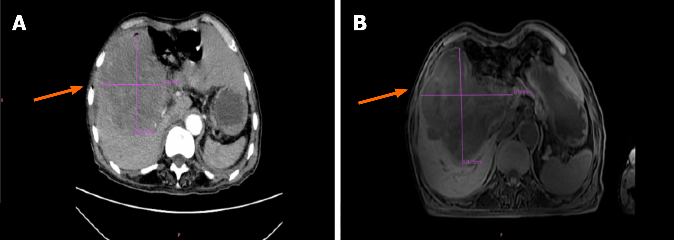
Figure 1 Preoperative imaging.
A: Abdominal contrast-enhanced computed tomography showing a large, ill-defined heterogeneous mass (approximately 12.0 cm × 10.9 cm × 15.9 cm) extending from the subphrenic region to the gallbladder fossa and portacaval space (orange arrows), with infiltration of nearby liver, duodenum, and pancreatic head; B: Abdominal magnetic resonance imaging revealing a corresponding heterogeneous solid mass (measuring approximately 13.3 cm × 12.4 cm on axial imaging) in the right subphrenic/gallbladder fossa area (orange arrows).
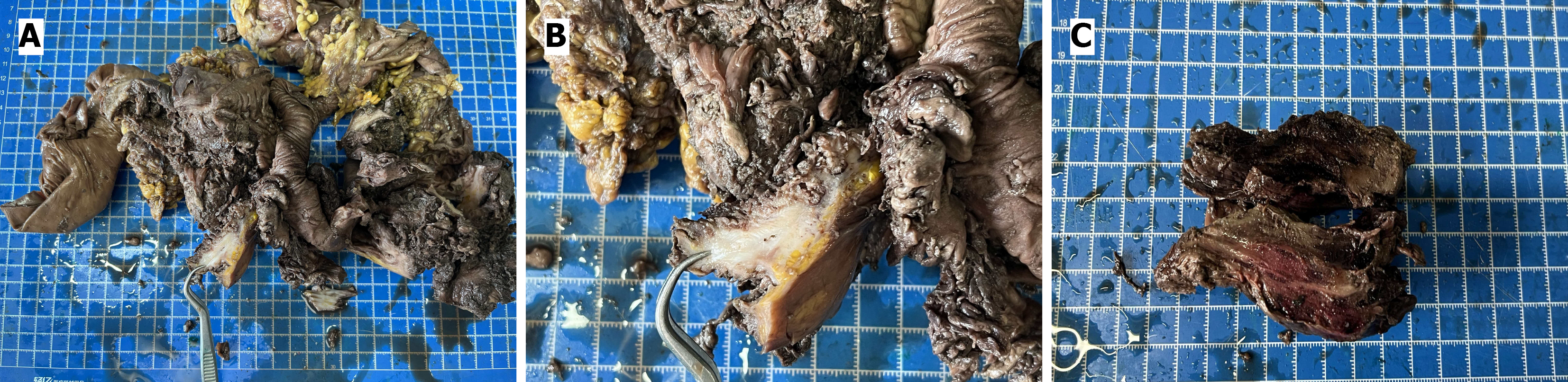
Figure 2 Gross photograph.
A: Gross view of the resected colonic specimen; B: The cut surface of the tumor shows gray-white areas; C: The cut surface exhibits a fish-flesh-like appearance.
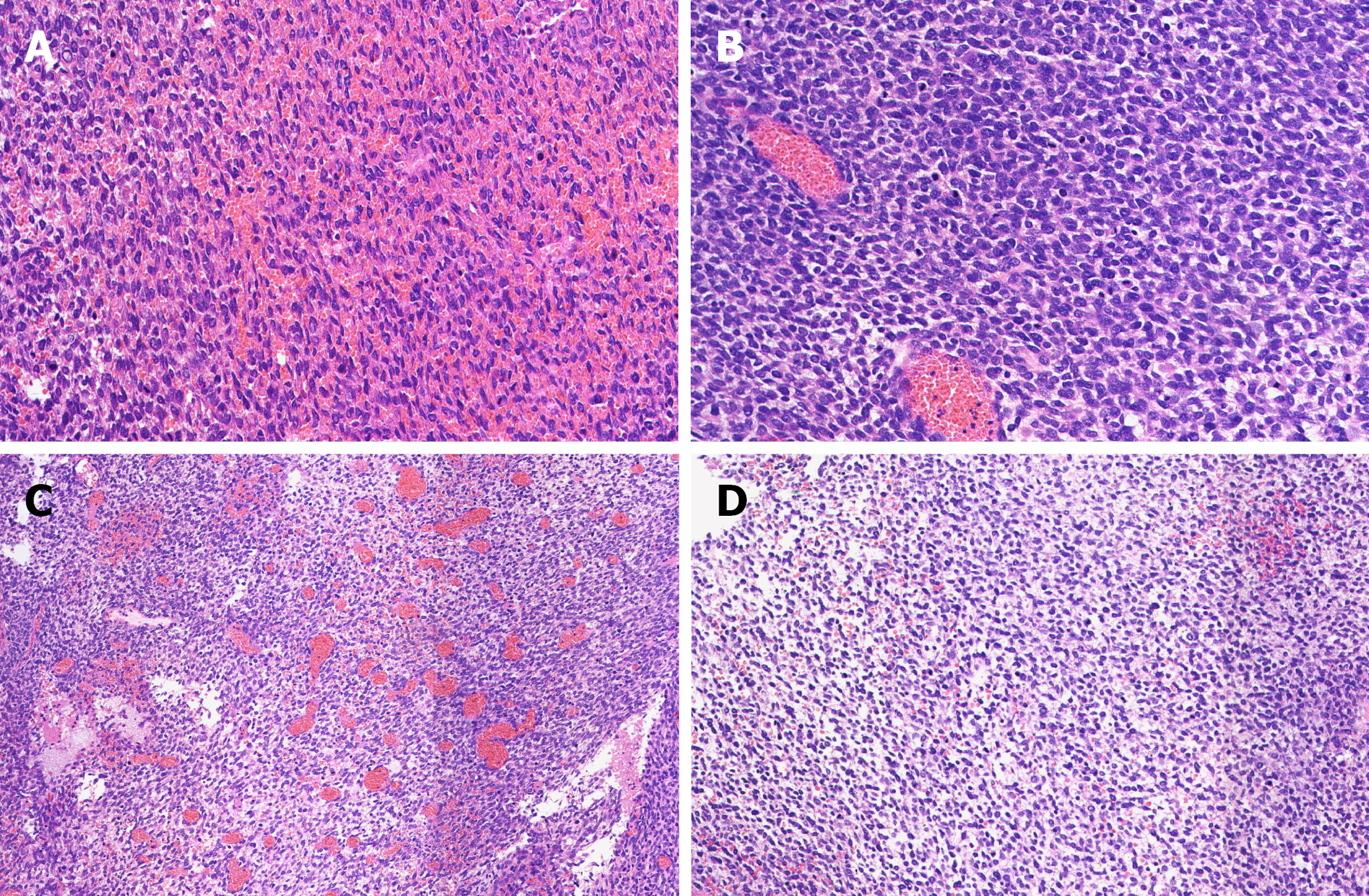
Figure 3 Histologic findings (A-D, all at × 200 magnification).
A: Fascicles of spindle-shaped tumor cells; B: Small round tumor cells; C: Tumor stroma with rich vascularity; D: Tumor stroma showing prominent myxoid change.
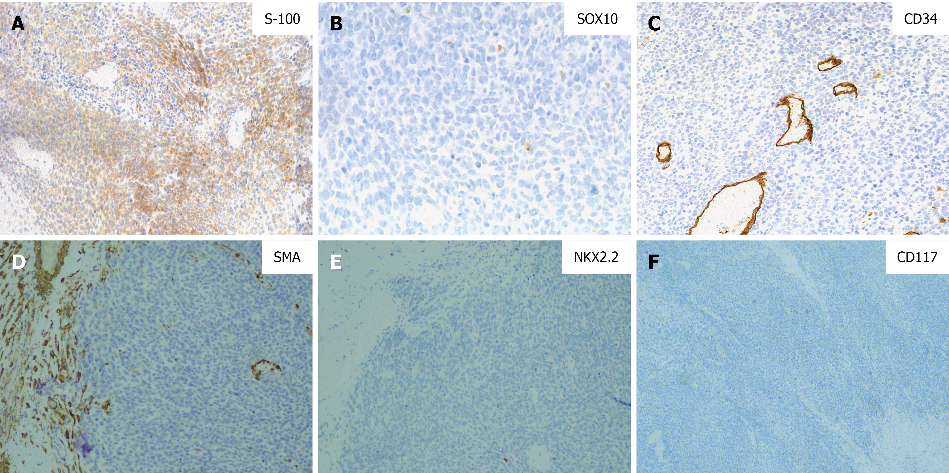
Figure 4 Immunohistochemical findings (A-F, all at × 100 magnification).
A: S-100 protein, focally positive; B: SOX10, negative; C: CD34 highlighting vascular structures only; D: SMA, negative; E: NKX2.2, negative; F: CD117, negative.
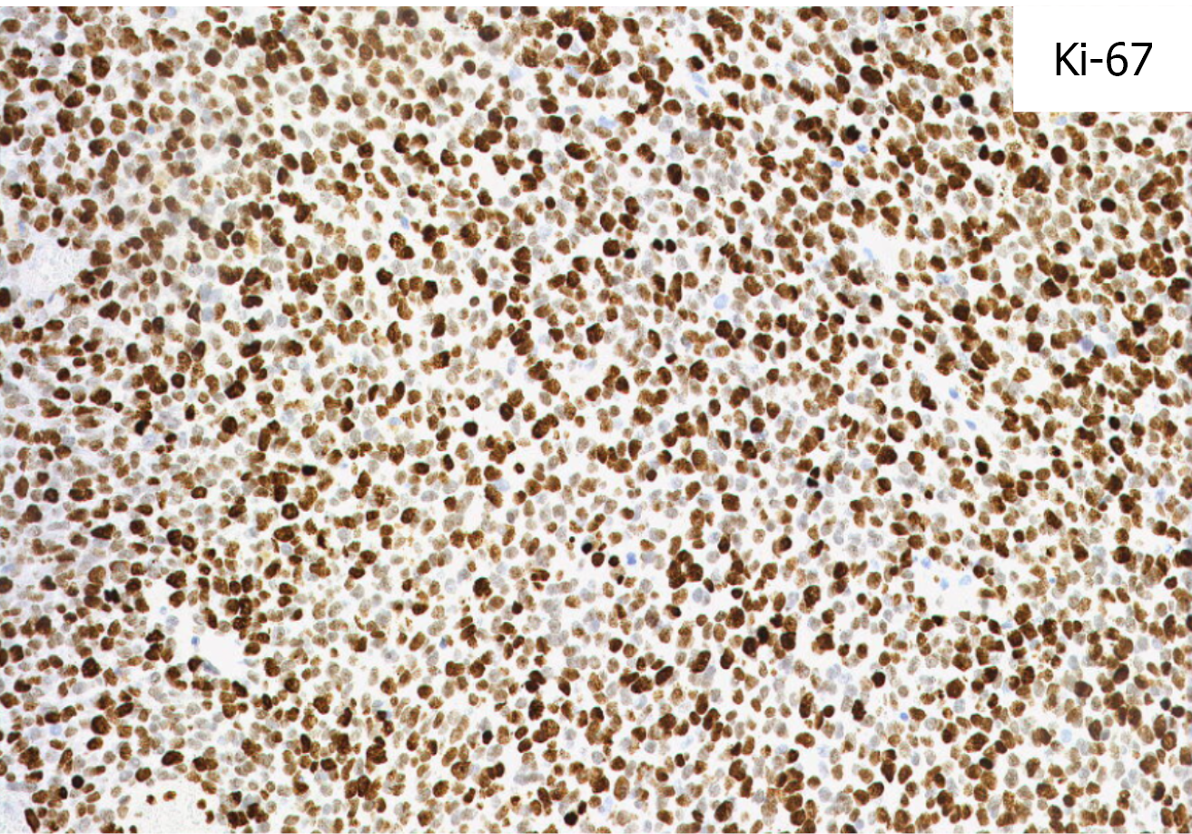
Figure 5
Immunohistochemical staining for Ki-67 showing a labeling index of approximately 90%.
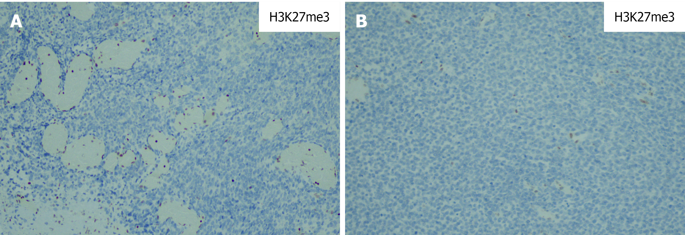
Figure 6 H3K27me3 immunohistochemical staining.
A: Retained positive nuclear staining in vascular endothelial cells serving as an internal control; B: Complete loss of H3K27me3 expression in all tumor cell nuclei.
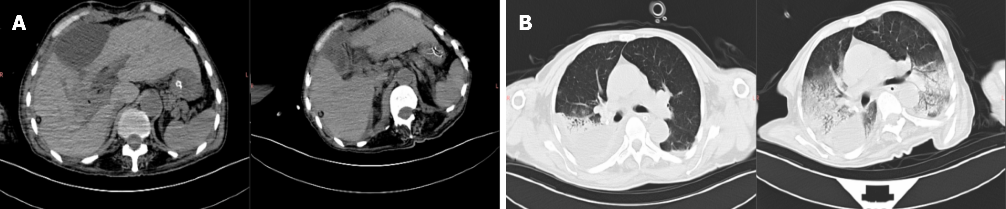
Figure 7 Postoperative imaging findings.
A: Imaging demonstrated moderate-to-large ascites; B: Massive pleural effusion was observed after surgery.
- Citation: Hu JY, He JX, Guo XJ, Zheng XY. Primary colonic malignant peripheral nerve sheath tumor with biphasic morphology: A case report. World J Gastrointest Oncol 2026; 18(10): 123676
- URL: https://www.wjgnet.com/1948-5204/full/v18/i10/123676.htm
- DOI: https://dx.doi.org/10.4251/wjgo.123676