















Copyright: ©Author(s) 2026.
World J Gastrointest Oncol. Oct 15, 2026; 18(10): 123534
Published online Oct 15, 2026. doi: 10.4251/wjgo.123534
Published online Oct 15, 2026. doi: 10.4251/wjgo.123534
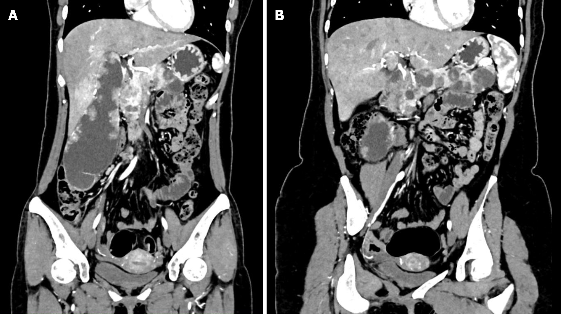
Figure 1 Contrast-enhanced computed tomography findings.
A: The gallbladder shows multiple soft-tissue lesions along the gallbladder wall and heterogeneous enhancement of a giant gallbladder; B: The pancreas shows multiple cystic-solid lesions distributed from the pancreatic head to the tail.
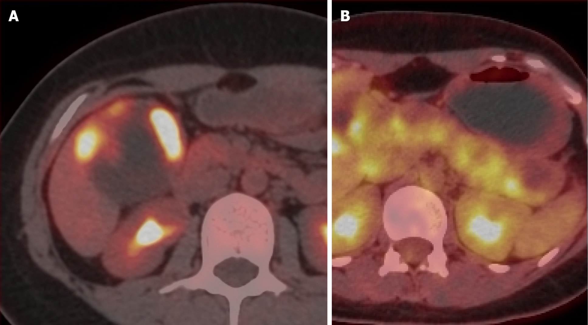
Figure 2 Positron emission tomography/computed tomography findings.
A: A cystic-solid mass in the gallbladder fossa shows a maximum standardized uptake value of 11.1; B: Multiple cystic-solid pancreatic masses show indistinct boundaries and heterogeneous uptake, with a maximum standardized uptake value of 7.9.
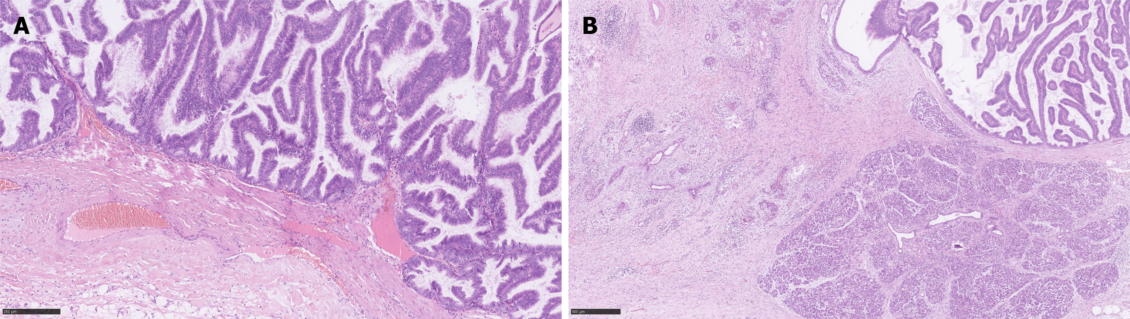
Figure 3 Histopathological features of gallbladder intracholecystic papillary-tubular neoplasm and pancreatic intraductal tubulopapillary neoplasm.
A: The gallbladder lesion shows an intraluminal papillary-tubular neoplastic proliferation with high-grade dysplasia; B: The pancreatic lesion shows predominantly intraductal epithelial neoplasm composed of tightly packed tubular and tubulopapillary structures with multifocal invasive carcinoma infiltrating the pancreatic parenchyma. Hematoxylin and eosin staining.
- Citation: He XX, Feng YL, Zhang ZW, Xu TM, Li JN. Synchronous intracholecystic papillary-tubular neoplasm and pancreatic intraductal tubulopapillary neoplasm: A case report. World J Gastrointest Oncol 2026; 18(10): 123534
- URL: https://www.wjgnet.com/1948-5204/full/v18/i10/123534.htm
- DOI: https://dx.doi.org/10.4251/wjgo.123534