Copyright
©The Author(s) 2025.
World J Gastrointest Oncol. Aug 15, 2025; 17(8): 110206
Published online Aug 15, 2025. doi: 10.4251/wjgo.v17.i8.110206
Published online Aug 15, 2025. doi: 10.4251/wjgo.v17.i8.110206
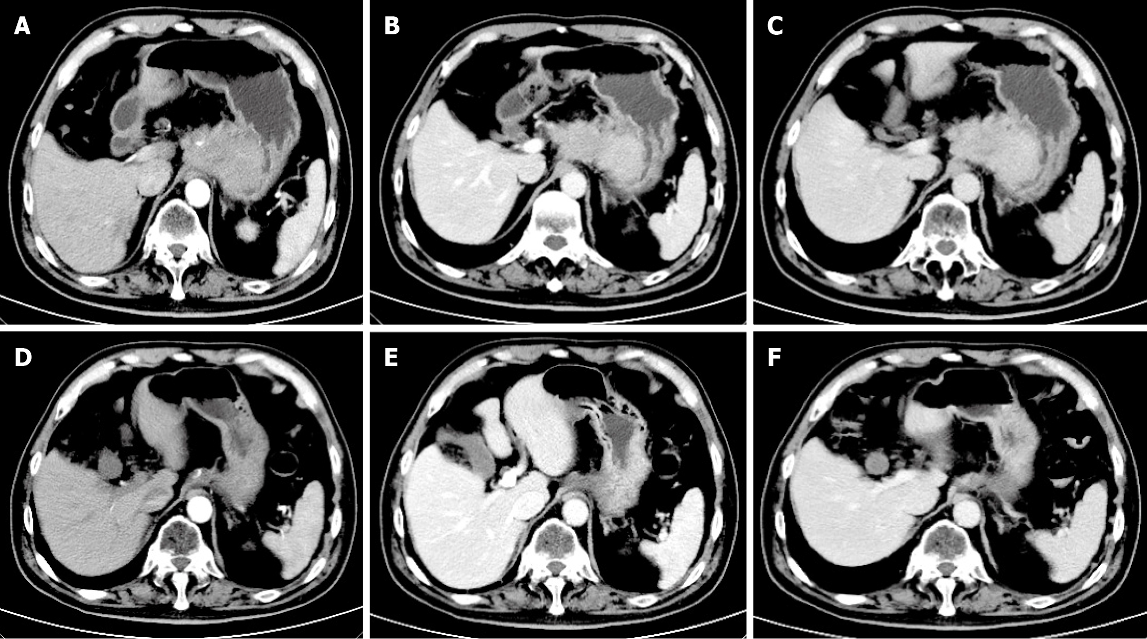
Figure 1 Abdominal computed tomography of case 1.
A: Arterial phase (before treatment); B: Venous phase (before treatment); C: Delayed phase (before treatment); D: Arterial phase (after 7 cycles of treatment); E: Venous phase (after 7 cycles of treatment); F: Delayed phase (after 7 cycles of treatment).
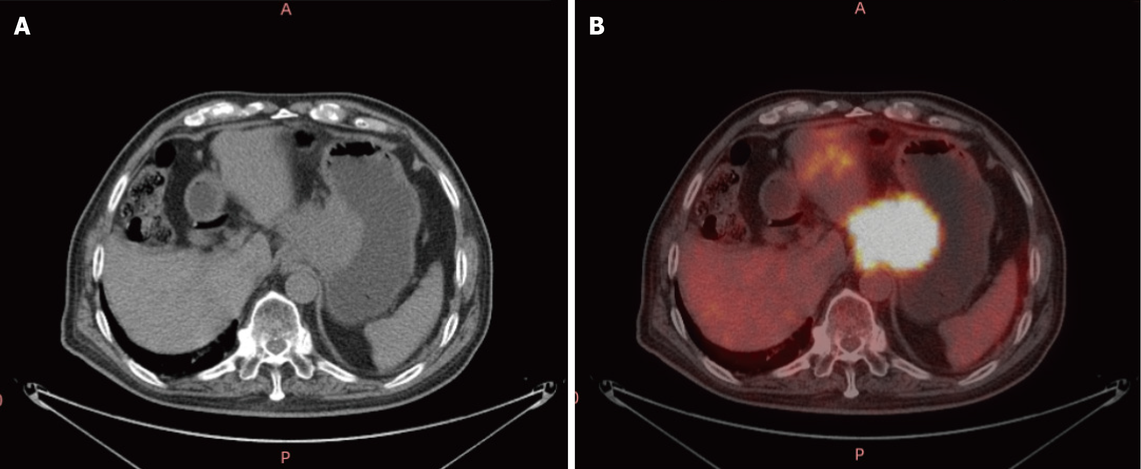
Figure 2 Positron emission tomography-computed tomography of case 1.
A: Abdominal window; B: Positron emission tomography window: Thickening of the gastric wall along the lesser curvature of the fundus and the presence of space-occupying lesions within the hepatogastric region.
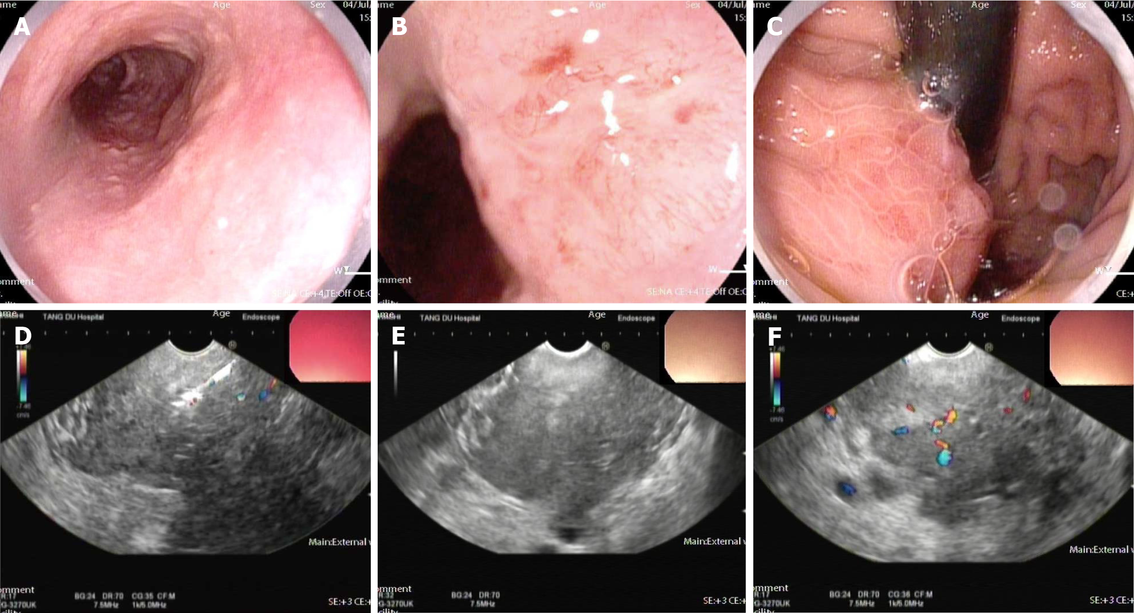
Figure 3 Gastroscopy and ultrasound gastroscopy of case 1.
A: Esophagus; B: Cardia; C: Fundus; D-F: Ultrasonic gastroscopy.
Figure 4 Pathology of case 1.
A: Esophagus; B: Cardia; C: Fundus: Squamous cell carcinoma hematoxylin and eosin stain 1:200.
- Citation: Wang HY, Song C, Ma J, Sun HQ, Yuan P, Liu ZX, Dou WJ. Challenges in the diagnosis of esophageal cancer with intramural gastric metastasis: Two case reports. World J Gastrointest Oncol 2025; 17(8): 110206
- URL: https://www.wjgnet.com/1948-5204/full/v17/i8/110206.htm
- DOI: https://dx.doi.org/10.4251/wjgo.v17.i8.110206


