Copyright
©The Author(s) 2025.
World J Gastrointest Oncol. Jun 15, 2025; 17(6): 106848
Published online Jun 15, 2025. doi: 10.4251/wjgo.v17.i6.106848
Published online Jun 15, 2025. doi: 10.4251/wjgo.v17.i6.106848
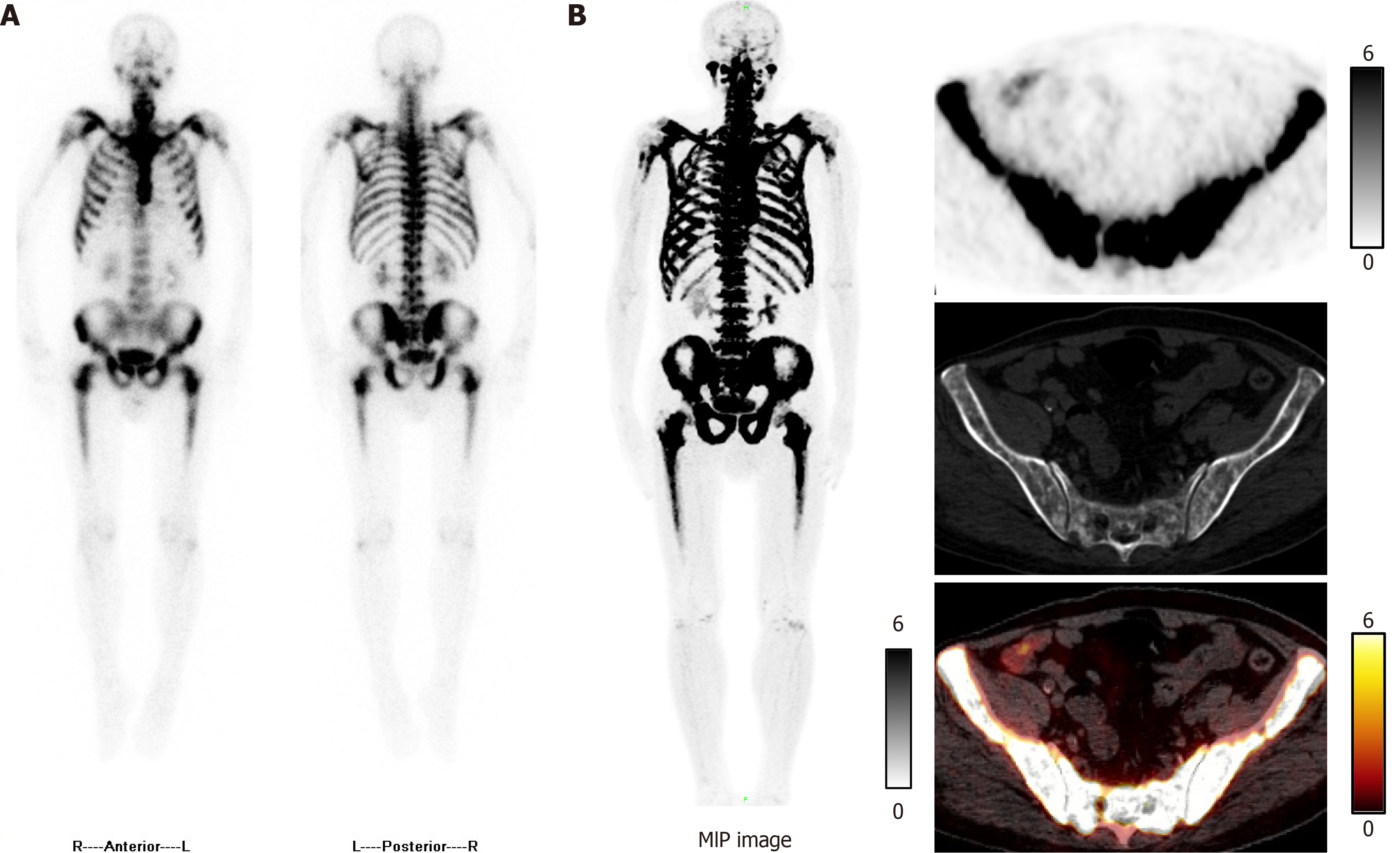
Figure 1 Technetium-99m methylene diphosphonate single-photon emission computed tomography and fluorine-18 sodium fluoride positron emission tomography/computed tomography images.
A: Whole-body bone scan showed diffuse increased technetium-99m methylene diphosphonate uptake throughout the skeleton; B: Symmetrical diffuse fluorine-18 sodium fluoride uptake was observed in the ribs, vertebrae, pelvis, and both femora. MIP: Maximum intensity projection.
Figure 2 Hematoxylin and eosin staining and immunohistochemical results of the patient’s bone tissue.
A: Pathology showed metastatic adenocarcinoma (400 ×); B: Immunochemistry of bone tissue showed positive staining for cytokeratin 7 (400 ×).
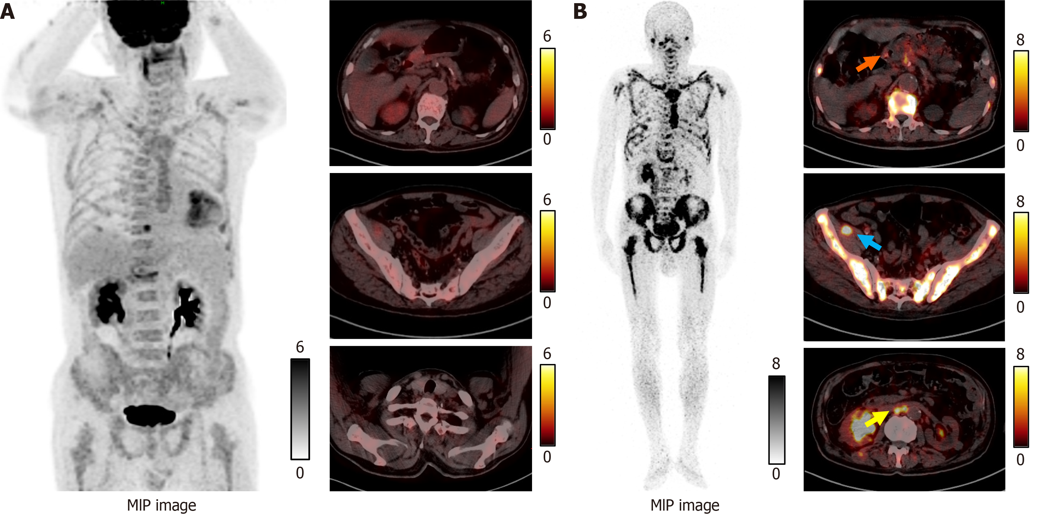
Figure 3 Fluorine-18 fluorodeoxyglucose and gallium-68-labeled fibroblast-activation protein inhibitor positron emission tomography/computed tomography images.
A: No obvious fluorine-18 fluorodeoxyglucose uptake was seen in the gastric antrum, pelvic cavity, or parathyroid region; B: Axial-fused gallium-68-labeled fibroblast-activation protein inhibitor positron emission tomography/computed tomography revealed increased uptake in the gastric antrum [maximum standardized uptake value (SUVmax): 5.52], right pelvic (SUVmax: 15.10), and abdominopelvic metastatic lesion (SUVmax: 10.50) (arrowhead). MIP: Maximum intensity projection.
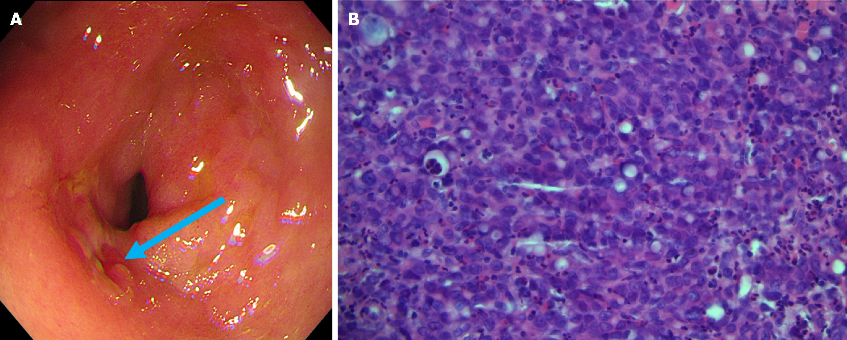
Figure 4 Patient’s gastroscopy and pathology images.
A: Gastric antral mucosa with scattered erythematous erosions, and a 1.0 cm erosion in the prepyloric area (blue arrow); B: Pathology showed the gastric antrum lesion to be consistent with poorly differentiated adenocarcinoma (hematoxylin and eosin 400 ×).
- Citation: Jin CB, Li YS, Zhang J, Wu J, Tao WJ. Extensive bone metastases from an occult gastric primary: A case report. World J Gastrointest Oncol 2025; 17(6): 106848
- URL: https://www.wjgnet.com/1948-5204/full/v17/i6/106848.htm
- DOI: https://dx.doi.org/10.4251/wjgo.v17.i6.106848


